 Movie
Movie Controller


+ Open data

- Basic information
| Entry | Database: PDB / ID: 7vdy | ||||||
|---|---|---|---|---|---|---|---|
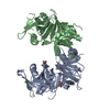
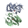
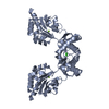
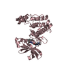
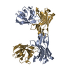
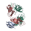
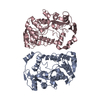
| Title | Crystal structure of O-ureidoserine racemase | ||||||
 Components Components | O-ureido-serine racemase | ||||||
 Keywords Keywords | ISOMERASE / racemase | ||||||
| Function / homology | O-ureido-serine racemase / diaminopimelate epimerase activity / Diaminopimelate epimerase, DapF / Diaminopimelate epimerase / racemase and epimerase activity, acting on amino acids and derivatives / L-lysine biosynthetic process via diaminopimelate / antibiotic biosynthetic process / cytoplasm / O-ureido-serine racemase Function and homology information Function and homology information | ||||||
| Biological species |  Streptomyces lavendulae (bacteria) Streptomyces lavendulae (bacteria) | ||||||
| Method |  X-RAY DIFFRACTION / SYNCHROTRON / MOLECULAR REPLACEMENT / Resolution: 2.12 Å X-RAY DIFFRACTION / SYNCHROTRON / MOLECULAR REPLACEMENT / Resolution: 2.12 Å | ||||||
 Authors Authors | Oda, K. / Matoba, Y. | ||||||
| Funding support | 1items
| ||||||
 Citation Citation | Journal: Proteins / Year: 2022 Title: Crystal structure of O-ureidoserine racemase found in the d-cycloserine biosynthetic pathway. Authors: Oda, K. / Sakaguchi, T. / Matoba, Y. | ||||||
| History |
|
- Structure visualization
| Structure viewer | Molecule: MolmilJmol/JSmol |
|---|
- Downloads & links
-Download
| PDBx/mmCIF format | 7vdy.cif.gz | 126.1 KB | Display | PDBx/mmCIF format |
|---|---|---|---|---|
| PDB format | pdb7vdy.ent.gz | 97.1 KB | Display | PDB format |
| PDBx/mmJSON format | 7vdy.json.gz | Tree view | PDBx/mmJSON format | |
| Others |  Other downloads Other downloads |
-Validation report
| Summary document | 7vdy_validation.pdf.gz | 454.4 KB | Display | wwPDB validaton report |
|---|---|---|---|---|
| Full document | 7vdy_full_validation.pdf.gz | 458.5 KB | Display | |
| Data in XML | 7vdy_validation.xml.gz | 25.8 KB | Display | |
| Data in CIF | 7vdy_validation.cif.gz | 37.6 KB | Display | |
| Arichive directory | https://data.pdbj.org/pub/pdb/validation_reports/vd/7vdyftp://data.pdbj.org/pub/pdb/validation_reports/vd/7vdy | HTTPS FTP |
-Related structure data
| Related structure data | 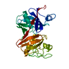 1gqzS S: Starting model for refinement |
|---|---|
| Similar structure data |


-Links
 PDBj
PDBj- Assembly
| Deposited unit | 
| ||||||||
|---|---|---|---|---|---|---|---|---|---|
| 1 |
| ||||||||
| Unit cell |
|
-Components
| #1: Protein | Mass: 31491.559 Da / Num. of mol.: 2 / Fragment: O-ureido-serine racemase Source method: isolated from a genetically manipulated source Source: (gene. exp.) Streptomyces lavendulae (bacteria) / Gene: dcsC, dapF / Plasmid: pET21 / Production host: #2: Chemical | ChemComp-SO4 /   Mass: 96.063 Da / Num. of mol.: 10 / Source method: obtained synthetically / Formula: SO4 Mass: 96.063 Da / Num. of mol.: 10 / Source method: obtained synthetically / Formula: SO4#3: Water | ChemComp-HOH / |  Mass: 18.015 Da / Num. of mol.: 385 / Source method: isolated from a natural source / Formula: H2O Mass: 18.015 Da / Num. of mol.: 385 / Source method: isolated from a natural source / Formula: H2OHas ligand of interest | N | |
|---|
-Experimental details
-Experiment
| Experiment | Method: X-RAY DIFFRACTION / Number of used crystals: 1 |
|---|
- Sample preparation
| Crystal | Density Matthews: 2.32 Å3/Da / Density % sol: 46.96 % / Mosaicity: 0.46 ° |
|---|---|
| Crystal grow | Temperature: 298 K / Method: vapor diffusion, sitting drop / pH: 6.5 / Details: 1.45 M ammonium sulfate, 10%(v/v) dioxane |
-Data collection
| Diffraction | Mean temperature: 100 K / Serial crystal experiment: N | ||||||||||||||||||||||||||||||
|---|---|---|---|---|---|---|---|---|---|---|---|---|---|---|---|---|---|---|---|---|---|---|---|---|---|---|---|---|---|---|---|
| Diffraction source | Source: SYNCHROTRON / Site: SPring-8  / Beamline: BL26B1 / Wavelength: 1 Å / Beamline: BL26B1 / Wavelength: 1 Å | ||||||||||||||||||||||||||||||
| Detector | Type: DECTRIS EIGER X 4M / Detector: PIXEL / Date: Jul 24, 2019 | ||||||||||||||||||||||||||||||
| Radiation | Protocol: SINGLE WAVELENGTH / Monochromatic (M) / Laue (L): M / Scattering type: x-ray | ||||||||||||||||||||||||||||||
| Radiation wavelength | Wavelength: 1 Å / Relative weight: 1 | ||||||||||||||||||||||||||||||
| Reflection | Resolution: 2.12→49.17 Å / Num. obs: 32385 / % possible obs: 99.4 % / Redundancy: 7 % / CC1/2: 0.994 / Rmerge(I) obs: 0.129 / Rpim(I) all: 0.052 / Rrim(I) all: 0.139 / Net I/σ(I): 9.6 / Num. measured all: 225765 / Scaling rejects: 305 | ||||||||||||||||||||||||||||||
| Reflection shell | Diffraction-ID: 1
|
- Processing
| Software |
| ||||||||||||||||||||||||||||||||||||||||||||||||||||||||||||||||||||||||||||||||||||
|---|---|---|---|---|---|---|---|---|---|---|---|---|---|---|---|---|---|---|---|---|---|---|---|---|---|---|---|---|---|---|---|---|---|---|---|---|---|---|---|---|---|---|---|---|---|---|---|---|---|---|---|---|---|---|---|---|---|---|---|---|---|---|---|---|---|---|---|---|---|---|---|---|---|---|---|---|---|---|---|---|---|---|---|---|---|
| Refinement | Method to determine structure: MOLECULAR REPLACEMENT Starting model: 1GQZ Resolution: 2.12→39.67 Å / SU ML: 0.22 / Cross valid method: THROUGHOUT / σ(F): 1.34 / Phase error: 23.07 / Stereochemistry target values: ML
| ||||||||||||||||||||||||||||||||||||||||||||||||||||||||||||||||||||||||||||||||||||
| Solvent computation | Shrinkage radii: 0.9 Å / VDW probe radii: 1.11 Å / Solvent model: FLAT BULK SOLVENT MODEL | ||||||||||||||||||||||||||||||||||||||||||||||||||||||||||||||||||||||||||||||||||||
| Displacement parameters | Biso max: 76.91 Å2 / Biso mean: 29.2221 Å2 / Biso min: 10.81 Å2 | ||||||||||||||||||||||||||||||||||||||||||||||||||||||||||||||||||||||||||||||||||||
| Refinement step | Cycle: final / Resolution: 2.12→39.67 Å
| ||||||||||||||||||||||||||||||||||||||||||||||||||||||||||||||||||||||||||||||||||||
| LS refinement shell | Refine-ID: X-RAY DIFFRACTION / Rfactor Rfree error: 0 / Total num. of bins used: 11
|
