 Movie
Movie Controller


+ Open data

- Basic information
| Entry | Database: PDB / ID: 7v6d | ||||||
|---|---|---|---|---|---|---|---|
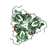
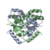
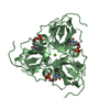
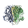
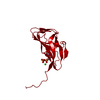
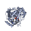
| Title | Structure of lipase B from Lasiodiplodia theobromae | ||||||
 Components Components | Lipase B | ||||||
 Keywords Keywords | HYDROLASE / alpha/beta-hydrolases | ||||||
| Function / homology | : / Alpha/Beta hydrolase fold, catalytic domain / Alpha/Beta hydrolase fold / Rossmann fold / 3-Layer(aba) Sandwich / metal ion binding / Alpha Beta / Lipase B Function and homology information Function and homology information | ||||||
| Biological species |  Lasiodiplodia theobromae (fungus) Lasiodiplodia theobromae (fungus) | ||||||
| Method |  X-RAY DIFFRACTION / SYNCHROTRON / MOLECULAR REPLACEMENT / molecular replacement / Resolution: 2.5 Å X-RAY DIFFRACTION / SYNCHROTRON / MOLECULAR REPLACEMENT / molecular replacement / Resolution: 2.5 Å | ||||||
 Authors Authors | Xue, B. / Zhang, H.F. / Nguyen, G.K.T. / Yew, W.S. | ||||||
| Funding support |  Singapore, 1items Singapore, 1items
| ||||||
 Citation Citation | Journal: Int J Mol Sci / Year: 2021 Title: A Novel Lipase from Lasiodiplodia theobromae Efficiently Hydrolyses C8-C10 Methyl Esters for the Preparation of Medium-Chain Triglycerides' Precursors. Authors: Ng, A.M.J. / Yang, R. / Zhang, H. / Xue, B. / Yew, W.S. / Nguyen, G.K.T. | ||||||
| History |
|
- Structure visualization
| Structure viewer | Molecule: MolmilJmol/JSmol |
|---|
- Downloads & links
-Download
| PDBx/mmCIF format | 7v6d.cif.gz | 151.1 KB | Display | PDBx/mmCIF format |
|---|---|---|---|---|
| PDB format | pdb7v6d.ent.gz | 115.2 KB | Display | PDB format |
| PDBx/mmJSON format | 7v6d.json.gz | Tree view | PDBx/mmJSON format | |
| Others |  Other downloads Other downloads |
-Validation report
| Summary document | 7v6d_validation.pdf.gz | 752 KB | Display | wwPDB validaton report |
|---|---|---|---|---|
| Full document | 7v6d_full_validation.pdf.gz | 756.9 KB | Display | |
| Data in XML | 7v6d_validation.xml.gz | 26.9 KB | Display | |
| Data in CIF | 7v6d_validation.cif.gz | 37.8 KB | Display | |
| Arichive directory | https://data.pdbj.org/pub/pdb/validation_reports/v6/7v6dftp://data.pdbj.org/pub/pdb/validation_reports/v6/7v6d | HTTPS FTP |
-Related structure data
| Related structure data | 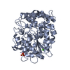 6idyS S: Starting model for refinement |
|---|---|
| Similar structure data |


-Links
 PDBj
PDBj- Assembly
| Deposited unit | 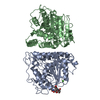
| ||||||||
|---|---|---|---|---|---|---|---|---|---|
| 1 | 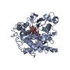
| ||||||||
| 2 | 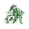
| ||||||||
| Unit cell |
|
-Components
| #1: Protein | Mass: 45076.785 Da / Num. of mol.: 2 Source method: isolated from a genetically manipulated source Source: (gene. exp.) Lasiodiplodia theobromae (fungus) / Gene: LIPB, DBV05_g2145 / Plasmid: pPIC9K / Production host: Komagataella pastoris (fungus) / Strain (production host): GS115 / References: UniProt: A0A5N5DNA6#2: Polysaccharide | 2-acetamido-2-deoxy-beta-D-glucopyranose-(1-4)-2-acetamido-2-deoxy-beta-D-glucopyranose | Source method: isolated from a genetically manipulated source #3: Chemical |   Mass: 40.078 Da / Num. of mol.: 2 / Source method: obtained synthetically / Formula: Ca Mass: 40.078 Da / Num. of mol.: 2 / Source method: obtained synthetically / Formula: Ca#4: Water | ChemComp-HOH / |  Mass: 18.015 Da / Num. of mol.: 118 / Source method: isolated from a natural source / Formula: H2O Mass: 18.015 Da / Num. of mol.: 118 / Source method: isolated from a natural source / Formula: H2OHas ligand of interest | N | Has protein modification | Y | |
|---|
-Experimental details
-Experiment
| Experiment | Method: X-RAY DIFFRACTION / Number of used crystals: 1 |
|---|
- Sample preparation
| Crystal | Density Matthews: 2.46 Å3/Da / Density % sol: 49.98 % / Mosaicity: 0.12 ° |
|---|---|
| Crystal grow | Temperature: 293 K / Method: vapor diffusion, sitting drop / pH: 4.6 / Details: 60% MPD, 100 mM NaOAc, pH 4.6, 10 mM CaCl2 |
-Data collection
| Diffraction | Mean temperature: 100 K / Serial crystal experiment: N | ||||||||||||||||||||||||||||||
|---|---|---|---|---|---|---|---|---|---|---|---|---|---|---|---|---|---|---|---|---|---|---|---|---|---|---|---|---|---|---|---|
| Diffraction source | Source: SYNCHROTRON / Site: Australian Synchrotron  / Beamline: MX1 / Wavelength: 0.95373 Å / Beamline: MX1 / Wavelength: 0.95373 Å | ||||||||||||||||||||||||||||||
| Detector | Type: DECTRIS EIGER X 9M / Detector: PIXEL / Date: Jun 9, 2020 | ||||||||||||||||||||||||||||||
| Radiation | Protocol: SINGLE WAVELENGTH / Monochromatic (M) / Laue (L): M / Scattering type: x-ray | ||||||||||||||||||||||||||||||
| Radiation wavelength | Wavelength: 0.95373 Å / Relative weight: 1 | ||||||||||||||||||||||||||||||
| Reflection | Resolution: 2.5→29.7 Å / Num. obs: 31233 / % possible obs: 99.9 % / Redundancy: 13.6 % / Biso Wilson estimate: 37.48 Å2 / CC1/2: 0.998 / Rmerge(I) obs: 0.191 / Rpim(I) all: 0.053 / Rrim(I) all: 0.198 / Net I/σ(I): 11.9 / Num. measured all: 425607 / Scaling rejects: 2 | ||||||||||||||||||||||||||||||
| Reflection shell | Diffraction-ID: 1
|
-Phasing
| Phasing | Method: molecular replacement |
|---|
- Processing
| Software |
| ||||||||||||||||||||||||||||||||||||||||||||||||||||||||||||||||||||||||
|---|---|---|---|---|---|---|---|---|---|---|---|---|---|---|---|---|---|---|---|---|---|---|---|---|---|---|---|---|---|---|---|---|---|---|---|---|---|---|---|---|---|---|---|---|---|---|---|---|---|---|---|---|---|---|---|---|---|---|---|---|---|---|---|---|---|---|---|---|---|---|---|---|---|
| Refinement | Method to determine structure: MOLECULAR REPLACEMENT Starting model: 6IDY Resolution: 2.5→29.7 Å / SU ML: 0.28 / Cross valid method: THROUGHOUT / σ(F): 1.34 / Phase error: 27.23 / Stereochemistry target values: ML
| ||||||||||||||||||||||||||||||||||||||||||||||||||||||||||||||||||||||||
| Solvent computation | Shrinkage radii: 0.9 Å / VDW probe radii: 1.11 Å / Solvent model: FLAT BULK SOLVENT MODEL | ||||||||||||||||||||||||||||||||||||||||||||||||||||||||||||||||||||||||
| Displacement parameters | Biso max: 98.1 Å2 / Biso mean: 43.7346 Å2 / Biso min: 20.06 Å2 | ||||||||||||||||||||||||||||||||||||||||||||||||||||||||||||||||||||||||
| Refinement step | Cycle: final / Resolution: 2.5→29.7 Å
| ||||||||||||||||||||||||||||||||||||||||||||||||||||||||||||||||||||||||
| LS refinement shell | Refine-ID: X-RAY DIFFRACTION / Rfactor Rfree error: 0 / Total num. of bins used: 11 / % reflection obs: 100 %
|
