 Movie
Movie Controller


+ Open data

- Basic information
| Entry | Database: PDB / ID: 7uxn | ||||||
|---|---|---|---|---|---|---|---|
| Title | Structure of PPIA in complex with FP29103, a Helicon Polypeptide | ||||||
 Components Components |
| ||||||
 Keywords Keywords | ISOMERASE / Complex / stapled | ||||||
| Function / homology |  Function and homology information Function and homology informationnegative regulation of protein K48-linked ubiquitination / regulation of apoptotic signaling pathway / cell adhesion molecule production / negative regulation of viral life cycle / lipid droplet organization / heparan sulfate binding / regulation of viral genome replication / virion binding / leukocyte chemotaxis / negative regulation of stress-activated MAPK cascade ...negative regulation of protein K48-linked ubiquitination / regulation of apoptotic signaling pathway / cell adhesion molecule production / negative regulation of viral life cycle / lipid droplet organization / heparan sulfate binding / regulation of viral genome replication / virion binding / leukocyte chemotaxis / negative regulation of stress-activated MAPK cascade / endothelial cell activation / Basigin interactions / protein peptidyl-prolyl isomerization / cyclosporin A binding / Minus-strand DNA synthesis / Plus-strand DNA synthesis / Uncoating of the HIV Virion / Early Phase of HIV Life Cycle / Integration of provirus / APOBEC3G mediated resistance to HIV-1 infection / viral release from host cell / negative regulation of protein phosphorylation / Calcineurin activates NFAT / Binding and entry of HIV virion / positive regulation of viral genome replication / negative regulation of oxidative stress-induced intrinsic apoptotic signaling pathway / activation of protein kinase B activity / neutrophil chemotaxis / Gene and protein expression by JAK-STAT signaling after Interleukin-12 stimulation / peptidyl-prolyl cis-trans isomerase activity / negative regulation of protein kinase activity / RNA polymerase II CTD heptapeptide repeat P3 isomerase activity / RNA polymerase II CTD heptapeptide repeat P6 isomerase activity / positive regulation of protein secretion / peptidylprolyl isomerase / Assembly Of The HIV Virion / Budding and maturation of HIV virion / platelet activation / platelet aggregation / neuron differentiation / positive regulation of NF-kappaB transcription factor activity / SARS-CoV-1 activates/modulates innate immune responses / integrin binding / unfolded protein binding / Platelet degranulation / protein folding / positive regulation of protein phosphorylation / cellular response to oxidative stress / secretory granule lumen / vesicle / ficolin-1-rich granule lumen / positive regulation of MAPK cascade / focal adhesion / apoptotic process / Neutrophil degranulation / protein-containing complex / extracellular space / RNA binding / extracellular exosome / extracellular region / nucleus / membrane / cytosol / cytoplasm Similarity search - Function | ||||||
| Biological species |  Homo sapiens (human) Homo sapiens (human)synthetic construct (others) | ||||||
| Method |  X-RAY DIFFRACTION / SYNCHROTRON / MOLECULAR REPLACEMENT / Resolution: 1.36 Å X-RAY DIFFRACTION / SYNCHROTRON / MOLECULAR REPLACEMENT / Resolution: 1.36 Å | ||||||
 Authors Authors | Li, K. / Agarwal, S. / Tokareva, O. / Thomson, T. / Tattersfield, H. / Wahl, S. / Verdine, G. / McGee, J. | ||||||
| Funding support |  United States, 1items United States, 1items
| ||||||
 Citation Citation | Journal: Proc.Natl.Acad.Sci.USA / Year: 2022 Title: De novo mapping of alpha-helix recognition sites on protein surfaces using unbiased libraries. Authors: Li, K. / Tokareva, O.S. / Thomson, T.M. / Wahl, S.C.T. / Travaline, T.L. / Ramirez, J.D. / Choudary, S.K. / Agarwal, S. / Walkup 4th, W.G. / Olsen, T.J. / Brennan, M.J. / Verdine, G.L. / McGee, J.H. | ||||||
| History |
|
- Structure visualization
| Structure viewer | Molecule: MolmilJmol/JSmol |
|---|
- Downloads & links
-Download
| PDBx/mmCIF format | 7uxn.cif.gz | 55.4 KB | Display | PDBx/mmCIF format |
|---|---|---|---|---|
| PDB format | pdb7uxn.ent.gz | 36.7 KB | Display | PDB format |
| PDBx/mmJSON format | 7uxn.json.gz | Tree view | PDBx/mmJSON format | |
| Others |  Other downloads Other downloads |
-Validation report
| Arichive directory | https://data.pdbj.org/pub/pdb/validation_reports/ux/7uxnftp://data.pdbj.org/pub/pdb/validation_reports/ux/7uxn | HTTPS FTP |
|---|
-Related structure data
| Related structure data | 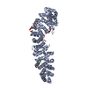 7uwiC 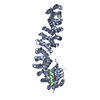 7uwoC 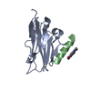 7ux5C  7uxiC  7uxjC 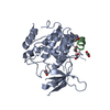 7uxkC  7uxmC  7uxoC  7uxpC  7uxqC  7uy2C  7uyjC  7uykC 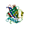 3k0nS C: citing same article ( S: Starting model for refinement |
|---|---|
| Similar structure data |
-Links
 PDBj
PDBj


- Assembly
| Deposited unit | 
| ||||||||||
|---|---|---|---|---|---|---|---|---|---|---|---|
| 1 |
| ||||||||||
| Unit cell |
|
-Components
| #1: Protein | Mass: 18093.555 Da / Num. of mol.: 1 Source method: isolated from a genetically manipulated source Source: (gene. exp.) Homo sapiens (human) / Gene: PPIA, CYPA / Production host:  |
|---|---|
| #2: Protein/peptide | Mass: 1342.569 Da / Num. of mol.: 1 / Source method: obtained synthetically / Details: Stapled peptide / Source: (synth.) synthetic construct (others) |
| #3: Chemical | ChemComp-WHL /   Mass: 192.214 Da / Num. of mol.: 1 / Source method: obtained synthetically / Formula: C10H12N2O2 Mass: 192.214 Da / Num. of mol.: 1 / Source method: obtained synthetically / Formula: C10H12N2O2 |
| #4: Water | ChemComp-HOH /  Mass: 18.015 Da / Num. of mol.: 197 / Source method: isolated from a natural source / Formula: H2O Mass: 18.015 Da / Num. of mol.: 197 / Source method: isolated from a natural source / Formula: H2O |
| Has ligand of interest | N |
| Has protein modification | Y |
-Experimental details
-Experiment
| Experiment | Method: X-RAY DIFFRACTION / Number of used crystals: 1 |
|---|
- Sample preparation
| Crystal | Density Matthews: 2.13 Å3/Da / Density % sol: 42.18 % |
|---|---|
| Crystal grow | Temperature: 291.15 K / Method: vapor diffusion, sitting drop Details: 0.2 M Sodium acetate, 0.1 M Sodium cacodylate pH 6.5, 30% w/v PEG 8000. |
-Data collection
| Diffraction | Mean temperature: 100 K / Serial crystal experiment: N |
|---|---|
| Diffraction source | Source: SYNCHROTRON / Site: ALS / Beamline: 5.0.1 / Wavelength: 0.97741 Å |
| Detector | Type: DECTRIS PILATUS3 6M / Detector: PIXEL / Date: May 23, 2021 |
| Radiation | Protocol: SINGLE WAVELENGTH / Monochromatic (M) / Laue (L): M / Scattering type: x-ray |
| Radiation wavelength | Wavelength: 0.97741 Å / Relative weight: 1 |
| Reflection | Resolution: 1.36→41.41 Å / Num. obs: 36286 / % possible obs: 99.73 % / Redundancy: 12.3 % / Biso Wilson estimate: 16.7 Å2 / CC1/2: 1 / Rmerge(I) obs: 0.044 / Net I/σ(I): 27.8 |
| Reflection shell | Resolution: 1.36→1.41 Å / Rmerge(I) obs: 0.951 / Num. unique obs: 3474 / CC1/2: 0.755 |
- Processing
| Software |
| |||||||||||||||||||||||||||||||||||||||||||||||||||||||||||||||||||||||||||||||||||||||||||||||||||||||||
|---|---|---|---|---|---|---|---|---|---|---|---|---|---|---|---|---|---|---|---|---|---|---|---|---|---|---|---|---|---|---|---|---|---|---|---|---|---|---|---|---|---|---|---|---|---|---|---|---|---|---|---|---|---|---|---|---|---|---|---|---|---|---|---|---|---|---|---|---|---|---|---|---|---|---|---|---|---|---|---|---|---|---|---|---|---|---|---|---|---|---|---|---|---|---|---|---|---|---|---|---|---|---|---|---|---|---|
| Refinement | Method to determine structure: MOLECULAR REPLACEMENT Starting model: 3K0N Resolution: 1.36→41.41 Å / SU ML: 0.15 / Cross valid method: THROUGHOUT / σ(F): 1.35 / Phase error: 21.74 / Stereochemistry target values: ML
| |||||||||||||||||||||||||||||||||||||||||||||||||||||||||||||||||||||||||||||||||||||||||||||||||||||||||
| Solvent computation | Shrinkage radii: 0.9 Å / VDW probe radii: 1.11 Å / Solvent model: FLAT BULK SOLVENT MODEL | |||||||||||||||||||||||||||||||||||||||||||||||||||||||||||||||||||||||||||||||||||||||||||||||||||||||||
| Displacement parameters | Biso max: 56.49 Å2 / Biso mean: 21.356 Å2 / Biso min: 10.49 Å2 | |||||||||||||||||||||||||||||||||||||||||||||||||||||||||||||||||||||||||||||||||||||||||||||||||||||||||
| Refinement step | Cycle: final / Resolution: 1.36→41.41 Å
| |||||||||||||||||||||||||||||||||||||||||||||||||||||||||||||||||||||||||||||||||||||||||||||||||||||||||
| LS refinement shell | Refine-ID: X-RAY DIFFRACTION / Rfactor Rfree error: 0 / Total num. of bins used: 14
|
