 Movie
Movie Controller


+ Open data

- Basic information
| Entry | Database: PDB / ID: 7uxk | ||||||
|---|---|---|---|---|---|---|---|
| Title | Structure of CDK2 in complex with FP24322, a Helicon Polypeptide | ||||||
 Components Components |
| ||||||
 Keywords Keywords | TRANSFERASE / Complex / stapled | ||||||
| Function / homology |  Function and homology information Function and homology informationcyclin A1-CDK2 complex / cyclin E2-CDK2 complex / cyclin E1-CDK2 complex / cyclin A2-CDK2 complex / positive regulation of DNA-templated DNA replication initiation / G2 Phase / Y chromosome / cyclin-dependent protein kinase activity / regulation of heterochromatin organization / Phosphorylation of proteins involved in G1/S transition by active Cyclin E:Cdk2 complexes ...cyclin A1-CDK2 complex / cyclin E2-CDK2 complex / cyclin E1-CDK2 complex / cyclin A2-CDK2 complex / positive regulation of DNA-templated DNA replication initiation / G2 Phase / Y chromosome / cyclin-dependent protein kinase activity / regulation of heterochromatin organization / Phosphorylation of proteins involved in G1/S transition by active Cyclin E:Cdk2 complexes / positive regulation of heterochromatin formation / p53-Dependent G1 DNA Damage Response / X chromosome / PTK6 Regulates Cell Cycle / regulation of anaphase-promoting complex-dependent catabolic process / Defective binding of RB1 mutants to E2F1,(E2F2, E2F3) / centriole replication / Regulation of APC/C activators between G1/S and early anaphase / telomere maintenance in response to DNA damage / centrosome duplication / G0 and Early G1 / Telomere Extension By Telomerase / Activation of the pre-replicative complex / cyclin-dependent kinase / cyclin-dependent protein serine/threonine kinase activity / TP53 Regulates Transcription of Genes Involved in G1 Cell Cycle Arrest / Activation of ATR in response to replication stress / Regulation of MITF-M-dependent genes involved in cell cycle and proliferation / Cajal body / Cyclin E associated events during G1/S transition / regulation of G2/M transition of mitotic cell cycle / Cyclin A:Cdk2-associated events at S phase entry / Cyclin A/B1/B2 associated events during G2/M transition / cyclin-dependent protein kinase holoenzyme complex / condensed chromosome / mitotic G1 DNA damage checkpoint signaling / cellular response to nitric oxide / negative regulation of protein localization to chromatin / regulation of mitotic cell cycle / post-translational protein modification / cyclin binding / positive regulation of DNA replication / male germ cell nucleus / potassium ion transport / peptidyl-serine phosphorylation / G1/S transition of mitotic cell cycle / meiotic cell cycle / DNA Damage/Telomere Stress Induced Senescence / Meiotic recombination / G2/M transition of mitotic cell cycle / CDK-mediated phosphorylation and removal of Cdc6 / cellular senescence / Transcriptional regulation of granulopoiesis / SCF(Skp2)-mediated degradation of p27/p21 / Orc1 removal from chromatin / Cyclin D associated events in G1 / Regulation of TP53 Degradation / nuclear envelope / Factors involved in megakaryocyte development and platelet production / regulation of gene expression / Processing of DNA double-strand break ends / Senescence-Associated Secretory Phenotype (SASP) / transcription regulator complex / Regulation of TP53 Activity through Phosphorylation / protein phosphorylation / Ras protein signal transduction / chromosome, telomeric region / DNA replication / endosome / chromatin remodeling / protein domain specific binding / protein serine kinase activity / cell division / DNA repair / protein serine/threonine kinase activity / centrosome / positive regulation of cell population proliferation / positive regulation of DNA-templated transcription / magnesium ion binding / negative regulation of transcription by RNA polymerase II / signal transduction / DNA-templated transcription / nucleoplasm / ATP binding / nucleus / cytosol / cytoplasm Similarity search - Function | ||||||
| Biological species |  Homo sapiens (human) Homo sapiens (human)synthetic construct (others) | ||||||
| Method |  X-RAY DIFFRACTION / SYNCHROTRON / MOLECULAR REPLACEMENT / Resolution: 2.63 Å X-RAY DIFFRACTION / SYNCHROTRON / MOLECULAR REPLACEMENT / Resolution: 2.63 Å | ||||||
 Authors Authors | Li, K. / Agarwal, S. / Tokareva, O. / Thomson, T. / Wahl, S. / Verdine, G. / McGee, J. | ||||||
| Funding support | 1items
| ||||||
 Citation Citation | Journal: Proc.Natl.Acad.Sci.USA / Year: 2022 Title: De novo mapping of alpha-helix recognition sites on protein surfaces using unbiased libraries. Authors: Li, K. / Tokareva, O.S. / Thomson, T.M. / Wahl, S.C.T. / Travaline, T.L. / Ramirez, J.D. / Choudary, S.K. / Agarwal, S. / Walkup 4th, W.G. / Olsen, T.J. / Brennan, M.J. / Verdine, G.L. / McGee, J.H. | ||||||
| History |
|
- Structure visualization
| Structure viewer | Molecule: MolmilJmol/JSmol |
|---|
- Downloads & links
-Download
| PDBx/mmCIF format | 7uxk.cif.gz | 68.2 KB | Display | PDBx/mmCIF format |
|---|---|---|---|---|
| PDB format | pdb7uxk.ent.gz | 47.5 KB | Display | PDB format |
| PDBx/mmJSON format | 7uxk.json.gz | Tree view | PDBx/mmJSON format | |
| Others |  Other downloads Other downloads |
-Validation report
| Arichive directory | https://data.pdbj.org/pub/pdb/validation_reports/ux/7uxkftp://data.pdbj.org/pub/pdb/validation_reports/ux/7uxk | HTTPS FTP |
|---|
-Related structure data
| Related structure data | 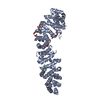 7uwiC 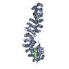 7uwoC 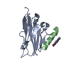 7ux5C  7uxiC  7uxjC  7uxmC  7uxnC  7uxoC  7uxpC  7uxqC  7uy2C  7uyjC  7uykC 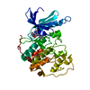 4kd1S C: citing same article ( S: Starting model for refinement |
|---|---|
| Similar structure data |
-Links
 PDBj
PDBj


- Assembly
| Deposited unit | 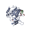
| ||||||||||
|---|---|---|---|---|---|---|---|---|---|---|---|
| 1 |
| ||||||||||
| Unit cell |
|
-Components
| #1: Protein | Mass: 34033.543 Da / Num. of mol.: 1 Source method: isolated from a genetically manipulated source Source: (gene. exp.) Homo sapiens (human) / Gene: CDK2, CDKN2 / Production host:  | ||||||
|---|---|---|---|---|---|---|---|
| #2: Protein/peptide | Mass: 1207.375 Da / Num. of mol.: 1 / Source method: obtained synthetically / Source: (synth.) synthetic construct (others) | ||||||
| #3: Chemical | ChemComp-EDO /   Mass: 62.068 Da / Num. of mol.: 4 / Source method: obtained synthetically / Formula: C2H6O2 Mass: 62.068 Da / Num. of mol.: 4 / Source method: obtained synthetically / Formula: C2H6O2#4: Chemical | ChemComp-WHL / |   Mass: 192.214 Da / Num. of mol.: 1 / Source method: obtained synthetically / Formula: C10H12N2O2 Mass: 192.214 Da / Num. of mol.: 1 / Source method: obtained synthetically / Formula: C10H12N2O2Has ligand of interest | N | Has protein modification | Y | |
-Experimental details
-Experiment
| Experiment | Method: X-RAY DIFFRACTION / Number of used crystals: 1 |
|---|
- Sample preparation
| Crystal | Density Matthews: 2.21 Å3/Da / Density % sol: 44.27 % |
|---|---|
| Crystal grow | Temperature: 291.15 K / Method: vapor diffusion, hanging drop Details: Crystallization buffer: 0.1 M BICINE pH 9.2, 2% v/v 1,4-Dioxane, 6% w/v PEG 20000 |
-Data collection
| Diffraction | Mean temperature: 100 K / Serial crystal experiment: N |
|---|---|
| Diffraction source | Source: SYNCHROTRON / Site: APS  / Beamline: 19-ID / Wavelength: 0.97861 Å / Beamline: 19-ID / Wavelength: 0.97861 Å |
| Detector | Type: DECTRIS PILATUS3 X 6M / Detector: PIXEL / Date: Mar 15, 2021 |
| Radiation | Protocol: SINGLE WAVELENGTH / Monochromatic (M) / Laue (L): M / Scattering type: x-ray |
| Radiation wavelength | Wavelength: 0.97861 Å / Relative weight: 1 |
| Reflection | Resolution: 2.63→46.53 Å / Num. obs: 9922 / % possible obs: 99.51 % / Redundancy: 16.1 % / Biso Wilson estimate: 61.21 Å2 / CC1/2: 0.999 / Rmerge(I) obs: 0.129 / Net I/σ(I): 16.1 |
| Reflection shell | Resolution: 2.63→2.72 Å / Num. unique obs: 938 / CC1/2: 0.734 |
- Processing
| Software |
| ||||||||||||||||||||||||||||
|---|---|---|---|---|---|---|---|---|---|---|---|---|---|---|---|---|---|---|---|---|---|---|---|---|---|---|---|---|---|
| Refinement | Method to determine structure: MOLECULAR REPLACEMENT Starting model: 4KD1 Resolution: 2.63→46.53 Å / SU ML: 0.41 / Cross valid method: THROUGHOUT / σ(F): 1.34 / Phase error: 33.43 / Stereochemistry target values: ML
| ||||||||||||||||||||||||||||
| Solvent computation | Shrinkage radii: 0.9 Å / VDW probe radii: 1.11 Å / Solvent model: FLAT BULK SOLVENT MODEL | ||||||||||||||||||||||||||||
| Displacement parameters | Biso max: 153.98 Å2 / Biso mean: 70 Å2 / Biso min: 37.13 Å2 | ||||||||||||||||||||||||||||
| Refinement step | Cycle: final / Resolution: 2.63→46.53 Å
| ||||||||||||||||||||||||||||
| LS refinement shell | Refine-ID: X-RAY DIFFRACTION / Rfactor Rfree error: 0 / Total num. of bins used: 3
|
