 Movie
Movie Controller


+ Open data

- Basic information
| Entry | Database: PDB / ID: 7uxp | ||||||
|---|---|---|---|---|---|---|---|
| Title | Structure of PDL1 in complex with FP28132, a Helicon Polypeptide | ||||||
 Components Components |
| ||||||
 Keywords Keywords | IMMUNE SYSTEM / Complex / stapled | ||||||
| Function / homology |  Function and homology information Function and homology informationnegative regulation of tumor necrosis factor superfamily cytokine production / positive regulation of activated CD8-positive, alpha-beta T cell apoptotic process / negative regulation of CD8-positive, alpha-beta T cell activation / negative regulation of T cell mediated immune response to tumor cell / negative regulation of CD4-positive, alpha-beta T cell proliferation / TRIF-dependent toll-like receptor signaling pathway / STAT3 nuclear events downstream of ALK signaling / negative regulation of interleukin-10 production / negative regulation of T cell activation / negative regulation of activated T cell proliferation ...negative regulation of tumor necrosis factor superfamily cytokine production / positive regulation of activated CD8-positive, alpha-beta T cell apoptotic process / negative regulation of CD8-positive, alpha-beta T cell activation / negative regulation of T cell mediated immune response to tumor cell / negative regulation of CD4-positive, alpha-beta T cell proliferation / TRIF-dependent toll-like receptor signaling pathway / STAT3 nuclear events downstream of ALK signaling / negative regulation of interleukin-10 production / negative regulation of T cell activation / negative regulation of activated T cell proliferation / negative regulation of type II interferon production / positive regulation of interleukin-10 production / Co-inhibition by PD-1 / negative regulation of T cell receptor signaling pathway / negative regulation of T cell proliferation / T cell costimulation / response to cytokine / positive regulation of T cell proliferation / recycling endosome membrane / actin cytoskeleton / cellular response to lipopolysaccharide / early endosome membrane / adaptive immune response / transcription coactivator activity / cell surface receptor signaling pathway / immune response / receptor ligand activity / external side of plasma membrane / signal transduction / extracellular exosome / nucleoplasm / plasma membrane Similarity search - Function | ||||||
| Biological species |  Homo sapiens (human) Homo sapiens (human)synthetic construct (others) | ||||||
| Method |  X-RAY DIFFRACTION / SYNCHROTRON / MOLECULAR REPLACEMENT / Resolution: 2.62 Å X-RAY DIFFRACTION / SYNCHROTRON / MOLECULAR REPLACEMENT / Resolution: 2.62 Å | ||||||
 Authors Authors | Li, K. / Agarwal, S. / Tokareva, O. / Thomson, T. / Travaline, T. / Tattersfield, H. / Wahl, S. / Verdine, G. / McGee, J. | ||||||
| Funding support |  United States, 1items United States, 1items
| ||||||
 Citation Citation | Journal: Proc.Natl.Acad.Sci.USA / Year: 2022 Title: De novo mapping of alpha-helix recognition sites on protein surfaces using unbiased libraries. Authors: Li, K. / Tokareva, O.S. / Thomson, T.M. / Wahl, S.C.T. / Travaline, T.L. / Ramirez, J.D. / Choudary, S.K. / Agarwal, S. / Walkup 4th, W.G. / Olsen, T.J. / Brennan, M.J. / Verdine, G.L. / McGee, J.H. | ||||||
| History |
|
- Structure visualization
| Structure viewer | Molecule: MolmilJmol/JSmol |
|---|
- Downloads & links
-Download
| PDBx/mmCIF format | 7uxp.cif.gz | 70.9 KB | Display | PDBx/mmCIF format |
|---|---|---|---|---|
| PDB format | pdb7uxp.ent.gz | 46.1 KB | Display | PDB format |
| PDBx/mmJSON format | 7uxp.json.gz | Tree view | PDBx/mmJSON format | |
| Others |  Other downloads Other downloads |
-Validation report
| Arichive directory | https://data.pdbj.org/pub/pdb/validation_reports/ux/7uxpftp://data.pdbj.org/pub/pdb/validation_reports/ux/7uxp | HTTPS FTP |
|---|
-Related structure data
| Related structure data | 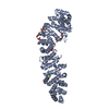 7uwiC 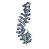 7uwoC 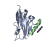 7ux5C  7uxiC  7uxjC 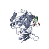 7uxkC  7uxmC  7uxnC  7uxoC  7uxqC  7uy2C  7uyjC  7uykC 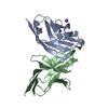 4zqkS C: citing same article ( S: Starting model for refinement |
|---|---|
| Similar structure data |
-Links
 PDBj
PDBj


- Assembly
| Deposited unit | 
| ||||||||||||
|---|---|---|---|---|---|---|---|---|---|---|---|---|---|
| 1 | 
| ||||||||||||
| 2 | 
| ||||||||||||
| Unit cell |
|
-Components
| #1: Protein | Mass: 14809.955 Da / Num. of mol.: 2 Source method: isolated from a genetically manipulated source Source: (gene. exp.) Homo sapiens (human) / Gene: CD274, B7H1, PDCD1L1, PDCD1LG1, PDL1 / Production host:  #2: Protein/peptide | Mass: 2015.248 Da / Num. of mol.: 2 / Source method: obtained synthetically / Source: (synth.) synthetic construct (others) #3: Chemical |   Mass: 16.023 Da / Num. of mol.: 2 / Source method: obtained synthetically / Formula: NH2 Mass: 16.023 Da / Num. of mol.: 2 / Source method: obtained synthetically / Formula: NH2#4: Chemical |   Mass: 192.214 Da / Num. of mol.: 2 / Source method: obtained synthetically / Formula: C10H12N2O2 Mass: 192.214 Da / Num. of mol.: 2 / Source method: obtained synthetically / Formula: C10H12N2O2#5: Water | ChemComp-HOH / |  Mass: 18.015 Da / Num. of mol.: 4 / Source method: isolated from a natural source / Formula: H2O Mass: 18.015 Da / Num. of mol.: 4 / Source method: isolated from a natural source / Formula: H2OHas ligand of interest | N | |
|---|
-Experimental details
-Experiment
| Experiment | Method: X-RAY DIFFRACTION / Number of used crystals: 1 |
|---|
- Sample preparation
| Crystal | Density Matthews: 2.31 Å3/Da / Density % sol: 46.86 % |
|---|---|
| Crystal grow | Temperature: 291.15 K / Method: vapor diffusion, sitting drop / Details: 0.1 M MES pH 6.5, 1.6 M Magnesium sulfate. |
-Data collection
| Diffraction | Mean temperature: 100 K / Serial crystal experiment: N |
|---|---|
| Diffraction source | Source: SYNCHROTRON / Site: Diamond  / Beamline: I03 / Wavelength: 0.97629 Å / Beamline: I03 / Wavelength: 0.97629 Å |
| Detector | Type: DECTRIS EIGER2 XE 16M / Detector: PIXEL / Date: Jul 2, 2021 |
| Radiation | Protocol: SINGLE WAVELENGTH / Monochromatic (M) / Laue (L): M / Scattering type: x-ray |
| Radiation wavelength | Wavelength: 0.97629 Å / Relative weight: 1 |
| Reflection | Resolution: 2.62→59.8 Å / Num. obs: 9672 / % possible obs: 99.9 % / Redundancy: 10 % / Biso Wilson estimate: 66.2 Å2 / CC1/2: 0.996 / Net I/σ(I): 11.7 |
| Reflection shell | Resolution: 2.62→2.71 Å / Num. unique obs: 953 / CC1/2: 0.841 |
- Processing
| Software |
| ||||||||||||||||||||||||||||
|---|---|---|---|---|---|---|---|---|---|---|---|---|---|---|---|---|---|---|---|---|---|---|---|---|---|---|---|---|---|
| Refinement | Method to determine structure: MOLECULAR REPLACEMENT Starting model: 4ZQK Resolution: 2.62→59.8 Å / SU ML: 0.505 / Cross valid method: FREE R-VALUE / σ(F): 1.38 / Phase error: 37.3348 Stereochemistry target values: GeoStd + Monomer Library + CDL v1.2
| ||||||||||||||||||||||||||||
| Solvent computation | Shrinkage radii: 0.9 Å / VDW probe radii: 1.11 Å / Solvent model: FLAT BULK SOLVENT MODEL | ||||||||||||||||||||||||||||
| Displacement parameters | Biso mean: 64.53 Å2 | ||||||||||||||||||||||||||||
| Refinement step | Cycle: LAST / Resolution: 2.62→59.8 Å
| ||||||||||||||||||||||||||||
| Refine LS restraints |
| ||||||||||||||||||||||||||||
| LS refinement shell |
|
