Movie
Movie Controller


+ Open data

- Basic information
| Entry | Database: PDB / ID: 7uws | |||||||||
|---|---|---|---|---|---|---|---|---|---|---|
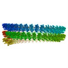
| Title | Atomic model of the partial VSV nucleocapsid | |||||||||
 Components Components |
| |||||||||
 Keywords Keywords | VIRAL PROTEIN/RNA / Vesicular stomatitis virus / Nucleocapsid / Matrix protein M / Nucleocapsid protein N / VIRAL PROTEIN-RNA complex | |||||||||
| Function / homology |  Function and homology information Function and homology informationRNA replication / host cell nuclear membrane / helical viral capsid / viral budding via host ESCRT complex / viral transcription / viral nucleocapsid / structural constituent of virion / host cell cytoplasm / ribonucleoprotein complex / viral envelope / RNA binding Similarity search - Function | |||||||||
| Biological species |  Vesicular stomatitis virus Vesicular stomatitis virus | |||||||||
| Method | ELECTRON MICROSCOPY / helical reconstruction / cryo EM / Resolution: 3.47 Å | |||||||||
 Authors Authors | Zhou, K. / Si, Z. / Ge, P. / Tsao, J. / Luo, M. / Zhou, Z.H. | |||||||||
| Funding support |  United States, 2items United States, 2items
| |||||||||
 Citation Citation | Journal: Nat Commun / Year: 2022 Title: Atomic model of vesicular stomatitis virus and mechanism of assembly. Authors: Kang Zhou / Zhu Si / Peng Ge / Jun Tsao / Ming Luo / Z Hong Zhou /  Abstract: Like other negative-strand RNA viruses (NSVs) such as influenza and rabies, vesicular stomatitis virus (VSV) has a three-layered organization: a layer of matrix protein (M) resides between the ...Like other negative-strand RNA viruses (NSVs) such as influenza and rabies, vesicular stomatitis virus (VSV) has a three-layered organization: a layer of matrix protein (M) resides between the glycoprotein (G)-studded membrane envelope and the nucleocapsid, which is composed of the nucleocapsid protein (N) and the encapsidated genomic RNA. Lack of in situ atomic structures of these viral components has limited mechanistic understanding of assembling the bullet-shaped virion. Here, by cryoEM and sub-particle reconstruction, we have determined the in situ structures of M and N inside VSV at 3.47 Å resolution. In the virion, N and M sites have a stoichiometry of 1:2. The in situ structures of both N and M differ from their crystal structures in their N-terminal segments and oligomerization loops. N-RNA, N-N, and N-M-M interactions govern the formation of the capsid. A double layer of M contributes to packaging of the helical nucleocapsid: the inner M (IM) joins neighboring turns of the N helix, while the outer M (OM) contacts G and the membrane envelope. The pseudo-crystalline organization of G is further mapped by cryoET. The mechanism of VSV assembly is delineated by the network interactions of these viral components. | |||||||||
| History |
|
- Structure visualization
| Structure viewer | Molecule: MolmilJmol/JSmol |
|---|
- Downloads & links
-Download
| PDBx/mmCIF format | 7uws.cif.gz | 848.5 KB | Display | PDBx/mmCIF format |
|---|---|---|---|---|
| PDB format | pdb7uws.ent.gz | 704.6 KB | Display | PDB format |
| PDBx/mmJSON format | 7uws.json.gz | Tree view | PDBx/mmJSON format | |
| Others |  Other downloads Other downloads |
-Validation report
| Arichive directory | https://data.pdbj.org/pub/pdb/validation_reports/uw/7uwsftp://data.pdbj.org/pub/pdb/validation_reports/uw/7uws | HTTPS FTP |
|---|
-Related structure data
| Related structure data | 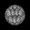 26841MC M: map data used to model this data C: citing same article ( |
|---|---|
| Similar structure data |
-Links
 PDBj
PDBj


- Assembly
| Deposited unit | 
|
|---|---|
| 1 |
|
-Components
| #1: Protein | Mass: 47463.949 Da / Num. of mol.: 7 / Source method: isolated from a natural source / Source: (natural) Vesicular stomatitis virus / Strain: San Juan / References: UniProt: P03521#2: RNA chain | | Mass: 116603.883 Da / Num. of mol.: 1 / Source method: isolated from a natural source / Source: (natural) Vesicular stomatitis virus / Strain: San Juan#3: Protein | Mass: 26228.188 Da / Num. of mol.: 12 / Source method: isolated from a natural source / Source: (natural) Vesicular stomatitis virus / References: UniProt: I7DGS2 |
|---|
-Experimental details
-Experiment
| Experiment | Method: ELECTRON MICROSCOPY |
|---|---|
| EM experiment | Aggregation state: HELICAL ARRAY / 3D reconstruction method: helical reconstruction |
- Sample preparation
| Component | Name: Vesicular stomatitis virus / Type: VIRUS / Entity ID: all / Source: NATURAL |
|---|---|
| Molecular weight | Value: 0.6 MDa / Experimental value: NO |
| Source (natural) | Organism: Vesicular stomatitis virus / Strain: San Juan |
| Details of virus | Empty: NO / Enveloped: YES / Isolate: STRAIN / Type: VIRION |
| Natural host | Organism: Homo sapiens |
| Virus shell | Name: Helical nucleocapsid / Diameter: 700 nm |
| Buffer solution | pH: 7.4 / Details: PBS buffer |
| Specimen | Embedding applied: NO / Shadowing applied: NO / Staining applied: NO / Vitrification applied: YES |
| Specimen support | Grid material: COPPER / Grid mesh size: 300 divisions/in. / Grid type: Quantifoil R1.2/1.3 |
| Vitrification | Instrument: FEI VITROBOT MARK IV / Cryogen name: ETHANE / Humidity: 100 % / Chamber temperature: 277 K |
- Electron microscopy imaging
| Experimental equipment |  Model: Titan Krios / Image courtesy: FEI Company |
|---|---|
| Microscopy | Model: FEI TITAN KRIOS |
| Electron gun | Electron source:  FIELD EMISSION GUN / Accelerating voltage: 300 kV / Illumination mode: FLOOD BEAM FIELD EMISSION GUN / Accelerating voltage: 300 kV / Illumination mode: FLOOD BEAM |
| Electron lens | Mode: BRIGHT FIELD / Nominal magnification: 105000 X / Nominal defocus max: 3000 nm / Nominal defocus min: 1500 nm / Cs: 2.7 mm / C2 aperture diameter: 75 µm / Alignment procedure: COMA FREE |
| Specimen holder | Cryogen: NITROGEN / Specimen holder model: FEI TITAN KRIOS AUTOGRID HOLDER |
| Image recording | Electron dose: 60 e/Å2 / Detector mode: COUNTING / Film or detector model: GATAN K2 SUMMIT (4k x 4k) / Num. of grids imaged: 1 / Num. of real images: 2008 |
| EM imaging optics | Energyfilter name: GIF Quantum LS / Energyfilter slit width: 20 eV |
| Image scans | Width: 3838 / Height: 3710 / Used frames/image: 2-44 |
- Processing
| EM software |
| ||||||||||||||||||||||||
|---|---|---|---|---|---|---|---|---|---|---|---|---|---|---|---|---|---|---|---|---|---|---|---|---|---|
| CTF correction | Type: PHASE FLIPPING AND AMPLITUDE CORRECTION | ||||||||||||||||||||||||
| Helical symmerty | Angular rotation/subunit: -10.14 ° / Axial rise/subunit: 1.42 Å / Axial symmetry: C1 | ||||||||||||||||||||||||
| Particle selection | Num. of particles selected: 7732 | ||||||||||||||||||||||||
| 3D reconstruction | Resolution: 3.47 Å / Resolution method: FSC 0.143 CUT-OFF / Num. of particles: 23207 Details: Additional sub-particle reconstruction was carried out after class3D of helices. Symmetry type: HELICAL | ||||||||||||||||||||||||
| Atomic model building | Protocol: AB INITIO MODEL / Space: REAL | ||||||||||||||||||||||||
| Atomic model building | 3D fitting-ID: 1 / Pdb chain-ID: A / Source name: PDB / Type: experimental model
|