 Movie
Movie Controller


+ Open data

- Basic information
| Entry | Database: PDB / ID: 2wyy | ||||||
|---|---|---|---|---|---|---|---|
| Title | CRYOEM MODEL OF THE VESICULAR STOMATITIS VIRUS | ||||||
 Components Components |
| ||||||
 Keywords Keywords | VIRUS / RNA / NSRV / HELIX / VIRION / VIRAL NUCLEOPROTEIN | ||||||
| Function / homology |  Function and homology information Function and homology informationRNA replication / helical viral capsid / viral transcription / viral nucleocapsid / host cell cytoplasm / ribonucleoprotein complex / RNA binding Similarity search - Function | ||||||
| Biological species |  VESICULAR STOMATITIS INDIANA VIRUS VESICULAR STOMATITIS INDIANA VIRUS | ||||||
| Method | ELECTRON MICROSCOPY / helical reconstruction / cryo EM / Resolution: 10.6 Å | ||||||
 Authors Authors | Ge, P. / Tsao, J. / Green, T.J. / Luo, M. / Zhou, Z.H. | ||||||
 Citation Citation | Journal: Science / Year: 2010 Title: Cryo-EM model of the bullet-shaped vesicular stomatitis virus. Authors: Peng Ge / Jun Tsao / Stan Schein / Todd J Green / Ming Luo / Z Hong Zhou /  Abstract: Vesicular stomatitis virus (VSV) is a bullet-shaped rhabdovirus and a model system of negative-strand RNA viruses. Through direct visualization by means of cryo-electron microscopy, we show that each ...Vesicular stomatitis virus (VSV) is a bullet-shaped rhabdovirus and a model system of negative-strand RNA viruses. Through direct visualization by means of cryo-electron microscopy, we show that each virion contains two nested, left-handed helices: an outer helix of matrix protein M and an inner helix of nucleoprotein N and RNA. M has a hub domain with four contact sites that link to neighboring M and N subunits, providing rigidity by clamping adjacent turns of the nucleocapsid. Side-by-side interactions between neighboring N subunits are critical for the nucleocapsid to form a bullet shape, and structure-based mutagenesis results support this description. Together, our data suggest a mechanism of VSV assembly in which the nucleocapsid spirals from the tip to become the helical trunk, both subsequently framed and rigidified by the M layer. | ||||||
| History |
|
- Structure visualization
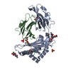
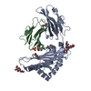
| Movie |
Movie viewer |
|---|---|
| Structure viewer | Molecule: MolmilJmol/JSmol |
- Downloads & links
-Download
| PDBx/mmCIF format | 2wyy.cif.gz | 763.5 KB | Display | PDBx/mmCIF format |
|---|---|---|---|---|
| PDB format | pdb2wyy.ent.gz | 627.8 KB | Display | PDB format |
| PDBx/mmJSON format | 2wyy.json.gz | Tree view | PDBx/mmJSON format | |
| Others |  Other downloads Other downloads |
-Validation report
| Arichive directory | https://data.pdbj.org/pub/pdb/validation_reports/wy/2wyyftp://data.pdbj.org/pub/pdb/validation_reports/wy/2wyy | HTTPS FTP |
|---|
-Related structure data
| Related structure data | 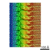 1663MC M: map data used to model this data C: citing same article ( |
|---|---|
| Similar structure data |
-Links
 PDBj
PDBj


- Assembly
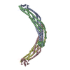
| Deposited unit | 
|
|---|---|
| 1 | x 15
|
| 2 |
|
| 3 | 
|
| Symmetry | Helical symmetry: (Circular symmetry: 1 / Dyad axis: no / N subunits divisor: 1 / Num. of operations: 15 / Rise per n subunits: 6.981 Å / Rotation per n subunits: -48 °) |
| Noncrystallographic symmetry (NCS) | NCS oper: (Code: given / Matrix: (0.669131, -0.743145), |
-Components
| #1: Protein | Mass: 47463.949 Da / Num. of mol.: 10 / Source method: isolated from a natural source / Source: (natural) VESICULAR STOMATITIS INDIANA VIRUS / Strain: SAN JUAN / References: UniProt: P03521#2: RNA chain | Mass: 13732.498 Da / Num. of mol.: 2 / Source method: isolated from a natural source / Source: (natural) VESICULAR STOMATITIS INDIANA VIRUS / Strain: SAN JUAN |
|---|
-Experimental details
-Experiment
| Experiment | Method: ELECTRON MICROSCOPY |
|---|---|
| EM experiment | Aggregation state: FILAMENT / 3D reconstruction method: helical reconstruction |
- Sample preparation
| Component | Name: VSV (INDIANA) / Type: VIRUS |
|---|---|
| Buffer solution | Name: 100MM NACL, 10NM TRIS, 1MM EDTA / pH: 7.4 / Details: 100MM NACL, 10NM TRIS, 1MM EDTA |
| Specimen | Embedding applied: NO / Shadowing applied: NO / Staining applied: NO / Vitrification applied: YES |
| Vitrification | Instrument: HOMEMADE PLUNGER / Cryogen name: ETHANE Details: CRYOGEN - ETHANE, HUMIDITY - 50, TEMPERATURE - 80, INSTRUMENT - MANUAL PLUNGER, METHOD- MANUAL BLOT 1 SECOND BEFORE PLUNGING, DETAILS - VITRIFICATION CARRIED OUT IN OPEN ROOM. |
- Electron microscopy imaging
| Experimental equipment |  Model: Tecnai Polara / Image courtesy: FEI Company |
|---|---|
| Microscopy | Model: FEI POLARA 300 / Date: Jan 5, 2007 |
| Electron gun | Electron source:  FIELD EMISSION GUN / Accelerating voltage: 300 kV / Illumination mode: SPOT SCAN FIELD EMISSION GUN / Accelerating voltage: 300 kV / Illumination mode: SPOT SCAN |
| Electron lens | Mode: BRIGHT FIELD / Nominal magnification: 59000 X / Calibrated magnification: 98000 X / Nominal defocus max: 2500 nm / Nominal defocus min: 800 nm / Cs: 2 mm |
| Specimen holder | Temperature: 80 K |
| Image recording | Electron dose: 20 e/Å2 / Film or detector model: GENERIC TVIPS |
| Image scans | Num. digital images: 212 |
| Radiation wavelength | Relative weight: 1 |
- Processing
| EM software |
| ||||||||||||
|---|---|---|---|---|---|---|---|---|---|---|---|---|---|
| 3D reconstruction | Method: PROJECTION MATCHING / Resolution: 10.6 Å Details: SUBMISSION BASED ON EXPERIMENTAL DATA FROM EMDB EMD-1663. Symmetry type: HELICAL | ||||||||||||
| Atomic model building | Protocol: RIGID BODY FIT / Space: REAL / Target criteria: Cross-correlation coefficient Details: METHOD--RIGID BODY REFINEMENT PROTOCOL--RIGID BODY REFINEMENT | ||||||||||||
| Atomic model building | PDB-ID: 2WYY Accession code: 2WYY / Source name: PDB / Type: experimental model | ||||||||||||
| Refinement | Highest resolution: 10.6 Å | ||||||||||||
| Refinement step | Cycle: LAST / Highest resolution: 10 Å
|
