 Movie
Movie Controller


+ Open data

- Basic information
| Entry | Database: PDB / ID: 7un1 | |||||||||||||||||||||
|---|---|---|---|---|---|---|---|---|---|---|---|---|---|---|---|---|---|---|---|---|---|---|
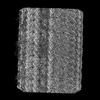
| Title | 8-nm repeat of the human sperm tip singlet microtubule | |||||||||||||||||||||
 Components Components |
| |||||||||||||||||||||
 Keywords Keywords | STRUCTURAL PROTEIN / cilia / microtubule / sperm / cell motility | |||||||||||||||||||||
| Function / homology |  Function and homology information Function and homology informationaxonemal microtubule doublet inner sheath / axoneme assembly / Post-chaperonin tubulin folding pathway / Cilium Assembly / axonemal microtubule / cytoskeleton-dependent intracellular transport / Carboxyterminal post-translational modifications of tubulin / Microtubule-dependent trafficking of connexons from Golgi to the plasma membrane / organelle transport along microtubule / forebrain morphogenesis ...axonemal microtubule doublet inner sheath / axoneme assembly / Post-chaperonin tubulin folding pathway / Cilium Assembly / axonemal microtubule / cytoskeleton-dependent intracellular transport / Carboxyterminal post-translational modifications of tubulin / Microtubule-dependent trafficking of connexons from Golgi to the plasma membrane / organelle transport along microtubule / forebrain morphogenesis / Sealing of the nuclear envelope (NE) by ESCRT-III / Intraflagellar transport / cerebellar cortex morphogenesis / glial cell differentiation / dentate gyrus development / neuron projection arborization / Formation of tubulin folding intermediates by CCT/TriC / flagellated sperm motility / Gap junction assembly / Kinesins / Prefoldin mediated transfer of substrate to CCT/TriC / pyramidal neuron differentiation / Assembly and cell surface presentation of NMDA receptors / COPI-independent Golgi-to-ER retrograde traffic / response to L-glutamate / centrosome cycle / COPI-dependent Golgi-to-ER retrograde traffic / natural killer cell mediated cytotoxicity / smoothened signaling pathway / ciliary base / regulation of synapse organization / startle response / motor behavior / Recycling pathway of L1 / microtubule polymerization / locomotory exploration behavior / response to tumor necrosis factor / MHC class I protein binding / axoneme / response to mechanical stimulus / sperm flagellum / microtubule-based process / intercellular bridge / RHO GTPases activate IQGAPs / Hedgehog 'off' state / COPI-mediated anterograde transport / cytoplasmic microtubule / Activation of AMPK downstream of NMDARs / condensed chromosome / Loss of Nlp from mitotic centrosomes / Loss of proteins required for interphase microtubule organization from the centrosome / Recruitment of mitotic centrosome proteins and complexes / MHC class II antigen presentation / Recruitment of NuMA to mitotic centrosomes / Anchoring of the basal body to the plasma membrane / HSP90 chaperone cycle for steroid hormone receptors (SHR) in the presence of ligand / Mitotic Prometaphase / homeostasis of number of cells within a tissue / EML4 and NUDC in mitotic spindle formation / cellular response to calcium ion / acrosomal vesicle / AURKA Activation by TPX2 / Resolution of Sister Chromatid Cohesion / Translocation of SLC2A4 (GLUT4) to the plasma membrane / adult locomotory behavior / neuromuscular junction / RHO GTPases Activate Formins / intracellular protein transport / cerebral cortex development / PKR-mediated signaling / synapse organization / visual learning / recycling endosome / structural constituent of cytoskeleton / microtubule cytoskeleton organization / neuron migration / memory / HCMV Early Events / calcium-dependent protein binding / Aggrephagy / cytoplasmic ribonucleoprotein granule / azurophil granule lumen / mitotic spindle / : / The role of GTSE1 in G2/M progression after G2 checkpoint / Separation of Sister Chromatids / Regulation of PLK1 Activity at G2/M Transition / mitotic cell cycle / double-stranded RNA binding / extracellular vesicle / microtubule cytoskeleton / neuron apoptotic process / microtubule binding / gene expression / cytoskeleton / microtubule / Hydrolases; Acting on acid anhydrides; Acting on GTP to facilitate cellular and subcellular movement / cilium / ciliary basal body / protein heterodimerization activity Similarity search - Function | |||||||||||||||||||||
| Biological species |  Homo sapiens (human) Homo sapiens (human) | |||||||||||||||||||||
| Method | ELECTRON MICROSCOPY / single particle reconstruction / cryo EM / Resolution: 6 Å | |||||||||||||||||||||
 Authors Authors | Gui, M. / Croft, J.T. / Zabeo, D. / Acharya, V. / Kollman, J.M. / Burgoyne, T. / Hoog, J.L. / Brown, A. | |||||||||||||||||||||
| Funding support |  United States, United States,  Sweden, 6items Sweden, 6items
| |||||||||||||||||||||
 Citation Citation | Journal: Proc Natl Acad Sci U S A / Year: 2022 Title: SPACA9 is a lumenal protein of human ciliary singlet and doublet microtubules. Authors: Miao Gui / Jacob T Croft / Davide Zabeo / Vajradhar Acharya / Justin M Kollman / Thomas Burgoyne / Johanna L Höög / Alan Brown /  Abstract: The cilium-centrosome complex contains triplet, doublet, and singlet microtubules. The lumenal surfaces of each microtubule within this diverse array are decorated by microtubule inner proteins ...The cilium-centrosome complex contains triplet, doublet, and singlet microtubules. The lumenal surfaces of each microtubule within this diverse array are decorated by microtubule inner proteins (MIPs). Here, we used single-particle cryo-electron microscopy methods to build atomic models of two types of human ciliary microtubule: the doublet microtubules of multiciliated respiratory cells and the distal singlet microtubules of monoflagellated human spermatozoa. We discover that SPACA9 is a polyspecific MIP capable of binding both microtubule types. SPACA9 forms intralumenal striations in the B tubule of respiratory doublet microtubules and noncontinuous spirals in sperm singlet microtubules. By acquiring new and reanalyzing previous cryo-electron tomography data, we show that SPACA9-like intralumenal striations are common features of different microtubule types in animal cilia. Our structures provide detailed references to help rationalize ciliopathy-causing mutations and position cryo-EM as a tool for the analysis of samples obtained directly from ciliopathy patients. | |||||||||||||||||||||
| History |
|
- Structure visualization
| Structure viewer | Molecule: MolmilJmol/JSmol |
|---|
- Downloads & links
-Download
| PDBx/mmCIF format | 7un1.cif.gz | 6.1 MB | Display | PDBx/mmCIF format |
|---|---|---|---|---|
| PDB format | pdb7un1.ent.gz | Display | PDB format | |
| PDBx/mmJSON format | 7un1.json.gz | Tree view | PDBx/mmJSON format | |
| Others |  Other downloads Other downloads |
-Validation report
| Arichive directory | https://data.pdbj.org/pub/pdb/validation_reports/un/7un1ftp://data.pdbj.org/pub/pdb/validation_reports/un/7un1 | HTTPS FTP |
|---|
-Related structure data
| Related structure data |  26611MC  7ungC M: map data used to model this data C: citing same article ( |
|---|---|
| Similar structure data |


-Links
 PDBj
PDBj


- Assembly
| Deposited unit | 
|
|---|---|
| 1 |
|
-Components
-Protein , 3 types, 109 molecules ABCDEFGHIJKLMNOPQRSTUVWXdefghi...
| #1: Protein | Mass: 25208.977 Da / Num. of mol.: 33 / Source method: isolated from a natural source / Source: (natural) Homo sapiens (human) / References: UniProt: Q96E40#2: Protein | Mass: 49877.824 Da / Num. of mol.: 38 / Source method: isolated from a natural source / Source: (natural) Homo sapiens (human) / References: UniProt: P68371#3: Protein | Mass: 50188.441 Da / Num. of mol.: 38 / Source method: isolated from a natural source / Source: (natural) Homo sapiens (human) / References: UniProt: Q71U36 |
|---|
-Non-polymers , 3 types, 114 molecules 


| #4: Chemical | ChemComp-GDP /  Type: RNA linking / Mass: 443.201 Da / Num. of mol.: 38 / Source method: obtained synthetically / Formula: C10H15N5O11P2 / Comment: GDP, energy-carrying molecule*YM Type: RNA linking / Mass: 443.201 Da / Num. of mol.: 38 / Source method: obtained synthetically / Formula: C10H15N5O11P2 / Comment: GDP, energy-carrying molecule*YM#5: Chemical | ChemComp-GTP /  Mass: 523.180 Da / Num. of mol.: 38 / Source method: obtained synthetically / Formula: C10H16N5O14P3 / Comment: GTP, energy-carrying molecule*YM Mass: 523.180 Da / Num. of mol.: 38 / Source method: obtained synthetically / Formula: C10H16N5O14P3 / Comment: GTP, energy-carrying molecule*YM#6: Chemical | ChemComp-MG /  Mass: 24.305 Da / Num. of mol.: 38 / Source method: obtained synthetically / Formula: Mg Mass: 24.305 Da / Num. of mol.: 38 / Source method: obtained synthetically / Formula: Mg |
|---|
-Details
| Has ligand of interest | N |
|---|
-Experimental details
-Experiment
| Experiment | Method: ELECTRON MICROSCOPY |
|---|---|
| EM experiment | Aggregation state: FILAMENT / 3D reconstruction method: single particle reconstruction |
- Sample preparation
| Component | Name: Singlet microtubule and associated SPACA9 / Type: COMPLEX / Entity ID: #1-#3 / Source: NATURAL |
|---|---|
| Molecular weight | Experimental value: NO |
| Source (natural) | Organism: Homo sapiens (human) |
| Buffer solution | pH: 7.4 |
| Specimen | Embedding applied: NO / Shadowing applied: NO / Staining applied: NO / Vitrification applied: YES / Details: Filaments |
| Specimen support | Grid material: COPPER / Grid mesh size: 200 divisions/in. / Grid type: EMS Lacey Carbon |
| Vitrification | Cryogen name: ETHANE |
- Electron microscopy imaging
| Experimental equipment |  Model: Titan Krios / Image courtesy: FEI Company |
|---|---|
| Microscopy | Model: FEI TITAN KRIOS |
| Electron gun | Electron source:  FIELD EMISSION GUN / Accelerating voltage: 300 kV / Illumination mode: FLOOD BEAM FIELD EMISSION GUN / Accelerating voltage: 300 kV / Illumination mode: FLOOD BEAM |
| Electron lens | Mode: BRIGHT FIELD / Nominal defocus max: 3000 nm / Nominal defocus min: 1000 nm |
| Image recording | Electron dose: 40 e/Å2 / Film or detector model: GATAN K3 BIOQUANTUM (6k x 4k) |
- Processing
| EM software |
| ||||||||||||||||
|---|---|---|---|---|---|---|---|---|---|---|---|---|---|---|---|---|---|
| CTF correction | Type: PHASE FLIPPING AND AMPLITUDE CORRECTION | ||||||||||||||||
| 3D reconstruction | Resolution: 6 Å / Resolution method: FSC 0.143 CUT-OFF / Num. of particles: 21990 / Symmetry type: POINT |
