 Movie
Movie Controller

[English] 日本語
 Yorodumi
- PDB-7ujr: Cocrystal structure of human CaMKII-alpha (CAMK2A)kinase domain a... -
+ Open data

- Basic information
| Entry | Database: PDB / ID: 7ujr | |||||||||
|---|---|---|---|---|---|---|---|---|---|---|
| Title | Cocrystal structure of human CaMKII-alpha (CAMK2A)kinase domain and GluN2B | |||||||||
 Components Components |
| |||||||||
 Keywords Keywords | TRANSFERASE / CaMKII / Kinase / Human / CAMK2A | |||||||||
| Function / homology |  Function and homology information Function and homology informationcalcium- and calmodulin-dependent protein kinase complex / regulation of endocannabinoid signaling pathway / Ca2+/calmodulin-dependent protein kinase / dendritic spine development / regulation of neuron migration / regulation of neurotransmitter secretion / Activated NTRK2 signals through FYN / excitatory chemical synaptic transmission / Synaptic adhesion-like molecules / Trafficking of AMPA receptors ...calcium- and calmodulin-dependent protein kinase complex / regulation of endocannabinoid signaling pathway / Ca2+/calmodulin-dependent protein kinase / dendritic spine development / regulation of neuron migration / regulation of neurotransmitter secretion / Activated NTRK2 signals through FYN / excitatory chemical synaptic transmission / Synaptic adhesion-like molecules / Trafficking of AMPA receptors / positive regulation of calcium ion transport / calcium/calmodulin-dependent protein kinase activity / regulation of mitochondrial membrane permeability involved in apoptotic process / Assembly and cell surface presentation of NMDA receptors / negative regulation of dendritic spine maintenance / Neurexins and neuroligins / regulation of monoatomic cation transmembrane transport / NMDA glutamate receptor activity / NMDA selective glutamate receptor complex / glutamate binding / glutamate receptor signaling pathway / CaMK IV-mediated phosphorylation of CREB / calcium ion transmembrane import into cytosol / protein heterotetramerization / glycine binding / Phase 0 - rapid depolarisation / ciliary transition zone / Dengue Virus-Host Interactions / Negative regulation of NMDA receptor-mediated neuronal transmission / Unblocking of NMDA receptors, glutamate binding and activation / negative regulation of ferroptosis / regulation of neuronal synaptic plasticity / Ion transport by P-type ATPases / Long-term potentiation / monoatomic cation transmembrane transport / HSF1-dependent transactivation / Regulation of MECP2 expression and activity / glutamate receptor binding / cellular response to interferon-beta / regulation of protein localization to plasma membrane / MECP2 regulates neuronal receptors and channels / Ion homeostasis / positive regulation of synaptic transmission, glutamatergic / ciliary tip / EPHB-mediated forward signaling / glutamate-gated calcium ion channel activity / positive regulation of cardiac muscle cell apoptotic process / ionotropic glutamate receptor signaling pathway / response to ischemia / ligand-gated monoatomic ion channel activity involved in regulation of presynaptic membrane potential / Ras activation upon Ca2+ influx through NMDA receptor / excitatory postsynaptic potential / positive regulation of excitatory postsynaptic potential / angiotensin-activated signaling pathway / positive regulation of receptor signaling pathway via JAK-STAT / synaptic membrane / synaptic transmission, glutamatergic / G1/S transition of mitotic cell cycle / transmitter-gated monoatomic ion channel activity involved in regulation of postsynaptic membrane potential / brain development / RAF activation / regulation of synaptic plasticity / cellular response to type II interferon / postsynaptic density membrane / Interferon gamma signaling / long-term synaptic potentiation / calcium ion transport / Signaling by RAF1 mutants / Signaling by moderate kinase activity BRAF mutants / Paradoxical activation of RAF signaling by kinase inactive BRAF / Signaling downstream of RAS mutants / endocytic vesicle membrane / Signaling by BRAF and RAF1 fusions / late endosome / amyloid-beta binding / RAF/MAP kinase cascade / Ca2+ pathway / chemical synaptic transmission / dendritic spine / response to ethanol / cytoskeleton / learning or memory / positive regulation of canonical NF-kappaB signal transduction / calmodulin binding / postsynaptic membrane / lysosome / cilium / neuron projection / postsynaptic density / protein serine kinase activity / protein serine/threonine kinase activity / dendrite / endoplasmic reticulum membrane / cell surface / protein homodimerization activity / mitochondrion / zinc ion binding / nucleoplasm / ATP binding / metal ion binding Similarity search - Function | |||||||||
| Biological species |  Homo sapiens (human) Homo sapiens (human) | |||||||||
| Method |  X-RAY DIFFRACTION / MOLECULAR REPLACEMENT / molecular replacement / Resolution: 1.95 Å X-RAY DIFFRACTION / MOLECULAR REPLACEMENT / molecular replacement / Resolution: 1.95 Å | |||||||||
 Authors Authors | Ozden, C. / Santos, N.J. / Stratton, M.M. / Garman, S.C. | |||||||||
| Funding support | 1items
| |||||||||
 Citation Citation | Journal: Cell Rep / Year: 2022 Title: CaMKII binds both substrates and activators at the active site. Authors: Ozden, C. / Sloutsky, R. / Mitsugi, T. / Santos, N. / Agnello, E. / Gaubitz, C. / Foster, J. / Lapinskas, E. / Esposito, E.A. / Saneyoshi, T. / Kelch, B.A. / Garman, S.C. / Hayashi, Y. / Stratton, M.M. | |||||||||
| History |
|
- Structure visualization
| Structure viewer | Molecule: MolmilJmol/JSmol |
|---|
- Downloads & links
-Download
| PDBx/mmCIF format | 7ujr.cif.gz | 75 KB | Display | PDBx/mmCIF format |
|---|---|---|---|---|
| PDB format | pdb7ujr.ent.gz | 51.9 KB | Display | PDB format |
| PDBx/mmJSON format | 7ujr.json.gz | Tree view | PDBx/mmJSON format | |
| Others |  Other downloads Other downloads |
-Validation report
| Arichive directory | https://data.pdbj.org/pub/pdb/validation_reports/uj/7ujrftp://data.pdbj.org/pub/pdb/validation_reports/uj/7ujr | HTTPS FTP |
|---|
-Related structure data
| Related structure data | 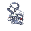 6x5gC 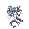 6x5qC 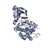 7kl0C 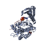 7kl1C 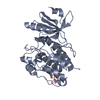 7uiqC 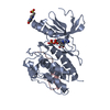 7uirC  7uisC 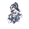 7ujpC 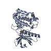 7ujqC 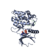 7ujsC 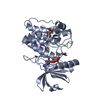 7ujtC 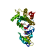 6vzkS C: citing same article ( S: Starting model for refinement |
|---|---|
| Similar structure data |
-Links
 PDBj
PDBj


- Assembly
| Deposited unit | 
| ||||||||
|---|---|---|---|---|---|---|---|---|---|
| 1 |
| ||||||||
| Unit cell |
|
-Components
| #1: Protein | Mass: 30549.074 Da / Num. of mol.: 1 / Mutation: Q223K Source method: isolated from a genetically manipulated source Source: (gene. exp.) Homo sapiens (human) / Gene: CAMK2A, CAMKA, KIAA0968 / Production host:  References: UniProt: Q9UQM7, Ca2+/calmodulin-dependent protein kinase | ||||||
|---|---|---|---|---|---|---|---|
| #2: Protein/peptide | Mass: 2754.155 Da / Num. of mol.: 1 / Source method: obtained synthetically / Source: (synth.) Homo sapiens (human) / References: UniProt: Q13224 | ||||||
| #3: Chemical |   Mass: 62.068 Da / Num. of mol.: 3 / Source method: obtained synthetically / Formula: C2H6O2 Mass: 62.068 Da / Num. of mol.: 3 / Source method: obtained synthetically / Formula: C2H6O2#4: Chemical | ChemComp-SO4 / |   Mass: 96.063 Da / Num. of mol.: 1 / Source method: obtained synthetically / Formula: SO4 Mass: 96.063 Da / Num. of mol.: 1 / Source method: obtained synthetically / Formula: SO4#5: Water | ChemComp-HOH / |  Mass: 18.015 Da / Num. of mol.: 93 / Source method: isolated from a natural source / Formula: H2O Mass: 18.015 Da / Num. of mol.: 93 / Source method: isolated from a natural source / Formula: H2OHas ligand of interest | N | |
-Experimental details
-Experiment
| Experiment | Method: X-RAY DIFFRACTION / Number of used crystals: 1 |
|---|
- Sample preparation
| Crystal | Density Matthews: 1.99 Å3/Da / Density % sol: 38.29 % |
|---|---|
| Crystal grow | Temperature: 277 K / Method: vapor diffusion, hanging drop / pH: 6.2 Details: 0.1M 1,3-bis(tris(hydroxymethyl)methylamino)propane, 0.1 M Ammonium sulfate, 20% PEG 3350, 5% Ethanol |
-Data collection
| Diffraction | Mean temperature: 100 K / Ambient temp details: 100 K thorughout the collection / Serial crystal experiment: N | |||||||||||||||||||||||||||||||||||||||||||||||||||||||||||||||||||||||||||||||||||||||||||||||||||||||||||||||||||||||||||||||||||||||||||||||||||||||||||||||||||||||||||||||||||||||||||||
|---|---|---|---|---|---|---|---|---|---|---|---|---|---|---|---|---|---|---|---|---|---|---|---|---|---|---|---|---|---|---|---|---|---|---|---|---|---|---|---|---|---|---|---|---|---|---|---|---|---|---|---|---|---|---|---|---|---|---|---|---|---|---|---|---|---|---|---|---|---|---|---|---|---|---|---|---|---|---|---|---|---|---|---|---|---|---|---|---|---|---|---|---|---|---|---|---|---|---|---|---|---|---|---|---|---|---|---|---|---|---|---|---|---|---|---|---|---|---|---|---|---|---|---|---|---|---|---|---|---|---|---|---|---|---|---|---|---|---|---|---|---|---|---|---|---|---|---|---|---|---|---|---|---|---|---|---|---|---|---|---|---|---|---|---|---|---|---|---|---|---|---|---|---|---|---|---|---|---|---|---|---|---|---|---|---|---|---|---|---|---|
| Diffraction source | Source: ROTATING ANODE / Type: RIGAKU MICROMAX-007 HF / Wavelength: 1.5418 Å | |||||||||||||||||||||||||||||||||||||||||||||||||||||||||||||||||||||||||||||||||||||||||||||||||||||||||||||||||||||||||||||||||||||||||||||||||||||||||||||||||||||||||||||||||||||||||||||
| Detector | Type: DECTRIS PILATUS3 R 200K-A / Detector: PIXEL / Date: Nov 18, 2019 / Details: Rigaku VariMax HF | |||||||||||||||||||||||||||||||||||||||||||||||||||||||||||||||||||||||||||||||||||||||||||||||||||||||||||||||||||||||||||||||||||||||||||||||||||||||||||||||||||||||||||||||||||||||||||||
| Radiation | Protocol: SINGLE WAVELENGTH / Monochromatic (M) / Laue (L): M / Scattering type: x-ray | |||||||||||||||||||||||||||||||||||||||||||||||||||||||||||||||||||||||||||||||||||||||||||||||||||||||||||||||||||||||||||||||||||||||||||||||||||||||||||||||||||||||||||||||||||||||||||||
| Radiation wavelength | Wavelength: 1.5418 Å / Relative weight: 1 | |||||||||||||||||||||||||||||||||||||||||||||||||||||||||||||||||||||||||||||||||||||||||||||||||||||||||||||||||||||||||||||||||||||||||||||||||||||||||||||||||||||||||||||||||||||||||||||
| Reflection | Resolution: 1.95→50 Å / Num. obs: 17848 / % possible obs: 93.8 % / Redundancy: 2.7 % / Rmerge(I) obs: 0.13 / Rpim(I) all: 0.094 / Rrim(I) all: 0.161 / Χ2: 5.482 / Net I/σ(I): 9.5 | |||||||||||||||||||||||||||||||||||||||||||||||||||||||||||||||||||||||||||||||||||||||||||||||||||||||||||||||||||||||||||||||||||||||||||||||||||||||||||||||||||||||||||||||||||||||||||||
| Reflection shell | Diffraction-ID: 1
|
-Phasing
| Phasing | Method: molecular replacement |
|---|
- Processing
| Software |
| ||||||||||||||||||||||||||||||||||||||||||||||||||||||||||||
|---|---|---|---|---|---|---|---|---|---|---|---|---|---|---|---|---|---|---|---|---|---|---|---|---|---|---|---|---|---|---|---|---|---|---|---|---|---|---|---|---|---|---|---|---|---|---|---|---|---|---|---|---|---|---|---|---|---|---|---|---|---|
| Refinement | Method to determine structure: MOLECULAR REPLACEMENT Starting model: 6vzk Resolution: 1.95→33.92 Å / Cor.coef. Fo:Fc: 0.946 / Cor.coef. Fo:Fc free: 0.916 / SU B: 4.667 / SU ML: 0.131 / Cross valid method: THROUGHOUT / σ(F): 0 / ESU R: 0.237 / ESU R Free: 0.19 / Stereochemistry target values: MAXIMUM LIKELIHOOD Details: HYDROGENS HAVE BEEN ADDED IN THE RIDING POSITIONS U VALUES : REFINED INDIVIDUALLY
| ||||||||||||||||||||||||||||||||||||||||||||||||||||||||||||
| Solvent computation | Ion probe radii: 0.8 Å / Shrinkage radii: 0.8 Å / VDW probe radii: 1.2 Å / Solvent model: MASK | ||||||||||||||||||||||||||||||||||||||||||||||||||||||||||||
| Displacement parameters | Biso max: 66.72 Å2 / Biso mean: 28.074 Å2 / Biso min: 19.02 Å2
| ||||||||||||||||||||||||||||||||||||||||||||||||||||||||||||
| Refinement step | Cycle: final / Resolution: 1.95→33.92 Å
| ||||||||||||||||||||||||||||||||||||||||||||||||||||||||||||
| Refine LS restraints |
| ||||||||||||||||||||||||||||||||||||||||||||||||||||||||||||
| LS refinement shell | Resolution: 1.951→2.002 Å / Rfactor Rfree error: 0 / Total num. of bins used: 20
|
