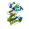
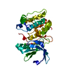
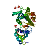
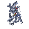
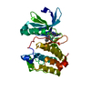
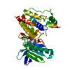
- PDB-6vzk: Crystal structure of human CaMKII-alpha (CAMK2A)kinase domain -
+
Open data
ID or keywords:
Loading...
-
Basic information
Entry
Database: PDB / ID: 6vzk
Title
Crystal structure of human CaMKII-alpha (CAMK2A)kinase domain
Components
Calcium/calmodulin-dependent protein kinase type II subunit alpha
Keywords
TRANSFERASE / CaMKII / Kinase / Human / CAMK2A
Function / homology
Function and homology information
calcium- and calmodulin-dependent protein kinase complex / regulation of endocannabinoid signaling pathway / Ca2+/calmodulin-dependent protein kinase / dendritic spine development / regulation of neuron migration / regulation of neurotransmitter secretion / Trafficking of AMPA receptors / positive regulation of calcium ion transport / calcium/calmodulin-dependent protein kinase activity / regulation of mitochondrial membrane permeability involved in apoptotic process ...calcium- and calmodulin-dependent protein kinase complex / regulation of endocannabinoid signaling pathway / Ca2+/calmodulin-dependent protein kinase / dendritic spine development / regulation of neuron migration / regulation of neurotransmitter secretion / Trafficking of AMPA receptors / positive regulation of calcium ion transport / calcium/calmodulin-dependent protein kinase activity / regulation of mitochondrial membrane permeability involved in apoptotic process / Assembly and cell surface presentation of NMDA receptors / CaMK IV-mediated phosphorylation of CREB / Phase 0 - rapid depolarisation / Dengue Virus-Host Interactions / Negative regulation of NMDA receptor-mediated neuronal transmission / Unblocking of NMDA receptors, glutamate binding and activation / negative regulation of ferroptosis / regulation of neuronal synaptic plasticity / Ion transport by P-type ATPases / Long-term potentiation / HSF1-dependent transactivation / Regulation of MECP2 expression and activity / glutamate receptor binding / cellular response to interferon-beta / regulation of protein localization to plasma membrane / Ion homeostasis / positive regulation of cardiac muscle cell apoptotic process / response to ischemia / Ras activation upon Ca2+ influx through NMDA receptor / angiotensin-activated signaling pathway / positive regulation of receptor signaling pathway via JAK-STAT / G1/S transition of mitotic cell cycle / RAF activation / cellular response to type II interferon / Interferon gamma signaling / long-term synaptic potentiation / calcium ion transport / Signaling by RAF1 mutants / Signaling by moderate kinase activity BRAF mutants / Paradoxical activation of RAF signaling by kinase inactive BRAF / Signaling downstream of RAS mutants / endocytic vesicle membrane / Signaling by BRAF and RAF1 fusions / RAF/MAP kinase cascade / Ca2+ pathway / dendritic spine / positive regulation of canonical NF-kappaB signal transduction / calmodulin binding / neuron projection / postsynaptic density / protein serine kinase activity / protein serine/threonine kinase activity / protein homodimerization activity / mitochondrion / nucleoplasm / ATP binding / metal ion binding / identical protein binding / nucleus / cytosol / cytoplasm Similarity search - Function
Calcium/calmodulin-dependent protein kinase II, association-domain / Calcium/calmodulin dependent protein kinase II association domain / NTF2-like domain superfamily / Phosphorylase Kinase; domain 1 / Phosphorylase Kinase; domain 1 / Serine/threonine-protein kinase, active site / Serine/Threonine protein kinases active-site signature. / Protein kinase domain / Serine/Threonine protein kinases, catalytic domain / Protein kinase, ATP binding site ...Calcium/calmodulin-dependent protein kinase II, association-domain / Calcium/calmodulin dependent protein kinase II association domain / NTF2-like domain superfamily / Phosphorylase Kinase; domain 1 / Phosphorylase Kinase; domain 1 / Serine/threonine-protein kinase, active site / Serine/Threonine protein kinases active-site signature. / Protein kinase domain / Serine/Threonine protein kinases, catalytic domain / Protein kinase, ATP binding site / Protein kinases ATP-binding region signature. / Protein kinase domain profile. / Protein kinase domain / Protein kinase-like domain superfamily / 2-Layer Sandwich / Alpha Beta Similarity search - Domain/homology
4'-HYDROXYCINNAMIC ACID / Calcium/calmodulin-dependent protein kinase type II subunit alpha Similarity search - Component
In the structure databanks used in Yorodumi, some data are registered as the other names, "COVID-19 virus" and "2019-nCoV". Here are the details of the virus and the list of structure data.
Jan 31, 2019. EMDB accession codes are about to change! (news from PDBe EMDB page)
EMDB accession codes are about to change! (news from PDBe EMDB page)
The allocation of 4 digits for EMDB accession codes will soon come to an end. Whilst these codes will remain in use, new EMDB accession codes will include an additional digit and will expand incrementally as the available range of codes is exhausted. The current 4-digit format prefixed with “EMD-” (i.e. EMD-XXXX) will advance to a 5-digit format (i.e. EMD-XXXXX), and so on. It is currently estimated that the 4-digit codes will be depleted around Spring 2019, at which point the 5-digit format will come into force.
The EM Navigator/Yorodumi systems omit the EMD- prefix.
Related info.:Q: What is EMD? / ID/Accession-code notation in Yorodumi/EM Navigator
Yorodumi is a browser for structure data from EMDB, PDB, SASBDB, etc.
This page is also the successor to EM Navigator detail page, and also detail information page/front-end page for Omokage search.
The word "yorodu" (or yorozu) is an old Japanese word meaning "ten thousand". "mi" (miru) is to see.
Related info.:EMDB / PDB / SASBDB / Comparison of 3 databanks / Yorodumi Search / Aug 31, 2016. New EM Navigator & Yorodumi / Yorodumi Papers / Jmol/JSmol / Function and homology information / Changes in new EM Navigator and Yorodumi
 Movie
Movie Controller


 Open data
 Basic information
 Components
 Keywords
 Function and homology information
 Homo sapiens (human)
 X-RAY DIFFRACTION /
  Authors
 United States, 1items
  Citation
 Structure visualization
 Downloads & links
 Other downloads
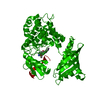


 PDBj
PDBj


 Assembly
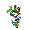


 Mass: 164.158 Da / Num. of mol.: 1 / Source method: obtained synthetically / Formula: C9H8O3
 Mass: 18.015 Da / Num. of mol.: 5 / Source method: isolated from a natural source / Formula: H2O
 Sample preparation
 Processing