 Movie
Movie Controller

[English] 日本語
 Yorodumi
- PDB-7uia: Crystal structure of BoNT/E receptor binding domain in complex wi... -
+ Open data

- Basic information
| Entry | Database: PDB / ID: 7uia | ||||||
|---|---|---|---|---|---|---|---|
| Title | Crystal structure of BoNT/E receptor binding domain in complex with SV2 and VHH | ||||||
 Components Components |
| ||||||
 Keywords Keywords | TOXIN / toxin-receptor complex | ||||||
| Function / homology |  Function and homology information Function and homology informationhost cell presynaptic membrane / host cell cytoplasmic vesicle / host cell cytosol / metalloendopeptidase activity / toxin activity / proteolysis / extracellular region / metal ion binding Similarity search - Function | ||||||
| Biological species |   Homo sapiens (human) Homo sapiens (human)  Clostridium botulinum (bacteria) Clostridium botulinum (bacteria) | ||||||
| Method |  X-RAY DIFFRACTION / SYNCHROTRON / MOLECULAR REPLACEMENT / Resolution: 2.59 Å X-RAY DIFFRACTION / SYNCHROTRON / MOLECULAR REPLACEMENT / Resolution: 2.59 Å | ||||||
 Authors Authors | Liu, Z. / Jin, R. / Chen, P. | ||||||
| Funding support |  United States, 1items United States, 1items
| ||||||
 Citation Citation | Journal: Nat Commun / Year: 2023 Title: Structural basis for botulinum neurotoxin E recognition of synaptic vesicle protein 2. Authors: Liu, Z. / Lee, P.G. / Krez, N. / Lam, K.H. / Liu, H. / Przykopanski, A. / Chen, P. / Yao, G. / Zhang, S. / Tremblay, J.M. / Perry, K. / Shoemaker, C.B. / Rummel, A. / Dong, M. / Jin, R. | ||||||
| History |
|
- Structure visualization
| Structure viewer | Molecule: MolmilJmol/JSmol |
|---|
- Downloads & links
-Download
| PDBx/mmCIF format | 7uia.cif.gz | 272 KB | Display | PDBx/mmCIF format |
|---|---|---|---|---|
| PDB format | pdb7uia.ent.gz | 214.3 KB | Display | PDB format |
| PDBx/mmJSON format | 7uia.json.gz | Tree view | PDBx/mmJSON format | |
| Others |  Other downloads Other downloads |
-Validation report
| Arichive directory | https://data.pdbj.org/pub/pdb/validation_reports/ui/7uiaftp://data.pdbj.org/pub/pdb/validation_reports/ui/7uia | HTTPS FTP |
|---|
-Related structure data
| Related structure data |  7uibC 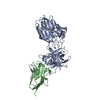 7uieC  3ffzS  5jlvS  6glwS S: Starting model for refinement C: citing same article ( |
|---|---|
| Similar structure data |
-Links
 PDBj
PDBj

- Assembly
| Deposited unit | 
| ||||||||
|---|---|---|---|---|---|---|---|---|---|
| 1 |
| ||||||||
| Unit cell |
|
-Components
-Protein , 2 types, 4 molecules CFDA
| #2: Protein | Mass: 12198.499 Da / Num. of mol.: 2 Source method: isolated from a genetically manipulated source Source: (gene. exp.) Homo sapiens (human)Production host: Mammalian expression vector BsrGI-MCS-pcDNA3.1 (others) #3: Protein | Mass: 47392.836 Da / Num. of mol.: 2 Source method: isolated from a genetically manipulated source Source: (gene. exp.) Clostridium botulinum (bacteria) / Production host: |
|---|
-Antibody , 1 types, 2 molecules BE
| #1: Antibody | Mass: 14390.949 Da / Num. of mol.: 2 Source method: isolated from a genetically manipulated source Source: (gene. exp.) Production host: Mammalian expression vector BsrGI-MCS-pcDNA3.1 (others) |
|---|
-Sugars , 2 types, 8 molecules 
| #4: Polysaccharide | Source method: isolated from a genetically manipulated source #5: Sugar | ChemComp-NAG /  Type: D-saccharide, beta linking / Mass: 221.208 Da / Num. of mol.: 6 Type: D-saccharide, beta linking / Mass: 221.208 Da / Num. of mol.: 6Source method: isolated from a genetically manipulated source Formula: C8H15NO6 / Feature type: SUBJECT OF INVESTIGATION |
|---|
-Non-polymers , 3 types, 167 molecules 


| #6: Chemical | ChemComp-SO4 /  Mass: 96.063 Da / Num. of mol.: 7 / Source method: obtained synthetically / Formula: SO4 Mass: 96.063 Da / Num. of mol.: 7 / Source method: obtained synthetically / Formula: SO4#7: Chemical | ChemComp-PEG / |  Mass: 106.120 Da / Num. of mol.: 1 / Source method: obtained synthetically / Formula: C4H10O3 Mass: 106.120 Da / Num. of mol.: 1 / Source method: obtained synthetically / Formula: C4H10O3#8: Water | ChemComp-HOH / | Mass: 18.015 Da / Num. of mol.: 159 / Source method: isolated from a natural source / Formula: H2O |
|---|
-Details
| Has ligand of interest | Y |
|---|---|
| Has protein modification | Y |
-Experimental details
-Experiment
| Experiment | Method: X-RAY DIFFRACTION / Number of used crystals: 1 |
|---|
- Sample preparation
| Crystal | Density Matthews: 2.85 Å3/Da / Density % sol: 56.91 % |
|---|---|
| Crystal grow | Temperature: 291 K / Method: vapor diffusion, hanging drop Details: 0.1 M HEPES, pH 7.5, 0.2 M potassium sulfate, 20% PEG 3350, and 5% (v/v) PEG 400 |
-Data collection
| Diffraction | Mean temperature: 100 K / Serial crystal experiment: N |
|---|---|
| Diffraction source | Source: SYNCHROTRON / Site: APS / Beamline: 24-ID-E / Wavelength: 0.9792 Å |
| Detector | Type: DECTRIS EIGER X 16M / Detector: PIXEL / Date: Nov 30, 2019 |
| Radiation | Protocol: SINGLE WAVELENGTH / Monochromatic (M) / Laue (L): M / Scattering type: x-ray |
| Radiation wavelength | Wavelength: 0.9792 Å / Relative weight: 1 |
| Reflection | Resolution: 2.59→137.16 Å / Num. obs: 52103 / % possible obs: 100 % / Redundancy: 12.1 % / CC1/2: 0.999 / Net I/σ(I): 19.9 |
| Reflection shell | Resolution: 2.59→2.67 Å / Num. unique obs: 4444 / CC1/2: 0.894 |
- Processing
| Software |
| ||||||||||||||||||||||||||||||||||||||||||||||||||||||||||||||||||||||||||||||||||||||||||||||||||||||||||||||||||||||||||||||||||||||||||||||||||||||||||||||||||||||||||||||||||||||
|---|---|---|---|---|---|---|---|---|---|---|---|---|---|---|---|---|---|---|---|---|---|---|---|---|---|---|---|---|---|---|---|---|---|---|---|---|---|---|---|---|---|---|---|---|---|---|---|---|---|---|---|---|---|---|---|---|---|---|---|---|---|---|---|---|---|---|---|---|---|---|---|---|---|---|---|---|---|---|---|---|---|---|---|---|---|---|---|---|---|---|---|---|---|---|---|---|---|---|---|---|---|---|---|---|---|---|---|---|---|---|---|---|---|---|---|---|---|---|---|---|---|---|---|---|---|---|---|---|---|---|---|---|---|---|---|---|---|---|---|---|---|---|---|---|---|---|---|---|---|---|---|---|---|---|---|---|---|---|---|---|---|---|---|---|---|---|---|---|---|---|---|---|---|---|---|---|---|---|---|---|---|---|---|
| Refinement | Method to determine structure: MOLECULAR REPLACEMENT Starting model: 3FFZ, 5JLV, 6GLW Resolution: 2.59→109.01 Å / Cor.coef. Fo:Fc: 0.932 / Cor.coef. Fo:Fc free: 0.919 / SU B: 11.9 / SU ML: 0.243 / Cross valid method: THROUGHOUT / ESU R: 0.711 / ESU R Free: 0.303 / Stereochemistry target values: MAXIMUM LIKELIHOOD / Details: HYDROGENS HAVE BEEN ADDED IN THE RIDING POSITIONS
| ||||||||||||||||||||||||||||||||||||||||||||||||||||||||||||||||||||||||||||||||||||||||||||||||||||||||||||||||||||||||||||||||||||||||||||||||||||||||||||||||||||||||||||||||||||||
| Solvent computation | Ion probe radii: 0.8 Å / Shrinkage radii: 0.8 Å / VDW probe radii: 1.2 Å / Solvent model: MASK | ||||||||||||||||||||||||||||||||||||||||||||||||||||||||||||||||||||||||||||||||||||||||||||||||||||||||||||||||||||||||||||||||||||||||||||||||||||||||||||||||||||||||||||||||||||||
| Displacement parameters | Biso mean: 51.741 Å2
| ||||||||||||||||||||||||||||||||||||||||||||||||||||||||||||||||||||||||||||||||||||||||||||||||||||||||||||||||||||||||||||||||||||||||||||||||||||||||||||||||||||||||||||||||||||||
| Refinement step | Cycle: 1 / Resolution: 2.59→109.01 Å
| ||||||||||||||||||||||||||||||||||||||||||||||||||||||||||||||||||||||||||||||||||||||||||||||||||||||||||||||||||||||||||||||||||||||||||||||||||||||||||||||||||||||||||||||||||||||
| Refine LS restraints |
|