National Institutes of Health/National Institute of Neurological Disorders and Stroke (NIH/NINDS)
DP1.NS111369
United States
Citation
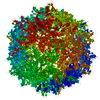
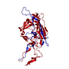
Journal: Mol Ther Methods Clin Dev / Year: 2022 Title: Structural basis of receptor usage by the engineered capsid AAV-PHP.eB. Authors: Seongmin Jang / Hao K Shen / Xiaozhe Ding / Timothy F Miles / Viviana Gradinaru / Abstract: Adeno-associated virus serotype 9 (AAV9) is a promising gene therapy vector for treating neurodegenerative diseases due to its ability to penetrate the blood-brain barrier. PHP.eB was engineered ...Adeno-associated virus serotype 9 (AAV9) is a promising gene therapy vector for treating neurodegenerative diseases due to its ability to penetrate the blood-brain barrier. PHP.eB was engineered from AAV9 by insertion of a 7-amino acid peptide and point mutation of neighboring residues, thereby enhancing potency in the central nervous system. Here, we report a 2.24-Å resolution cryo-electron microscopy structure of PHP.eB, revealing conformational differences from other 7-mer insertion capsid variants. In PHP.eB, the 7-mer loop adopts a bent conformation, mediated by an interaction between engineered lysine and aspartate residues. Further, we identify PKD2 as the main AAV receptor (AAVR) domain recognizing both AAV9 and PHP.eB and find that the PHP.eB 7-mer partially destabilizes this interaction. Analysis of previously reported AAV structures together with our pull-down data demonstrate that the 7-mer topology determined by the lysine-aspartate interaction dictates AAVR binding strength. Our results suggest that PHP.eB's altered tropism may arise from both an additional interaction with LY6A and weakening of its AAVR interaction. Changing the insertion length, but not sequence, modifies PKD2 binding affinity, suggesting that a steric clash impedes AAVR binding. This research suggests improved library designs for future AAV selections to identify non-LY6A-dependent vectors and modulate AAVR interaction strength.
Mass: 82188.609 Da / Num. of mol.: 1 Source method: isolated from a genetically manipulated source Source: (gene. exp.) Adeno-associated virus / Gene: cap / Production host: Homo sapiens (human) / References: UniProt: Q6JC40
-
Experimental details
-
Experiment
Experiment
Method: ELECTRON MICROSCOPY
EM experiment
Aggregation state: PARTICLE / 3D reconstruction method: single particle reconstruction
-
Sample preparation
Component
Name: Adeno-associated virus / Type: VIRUS / Details: AAV-PHP.eB: Engineered AAV from AAV9. / Entity ID: all / Source: RECOMBINANT
Source (natural)
Organism: Adeno-associated virus
Source (recombinant)
Organism: Homo sapiens (human) / Cell: AAVpro293T
Details of virus
Empty: NO / Enveloped: NO / Isolate: SEROTYPE / Type: VIRUS-LIKE PARTICLE
Buffer solution
pH: 7.4
Specimen
Embedding applied: NO / Shadowing applied: NO / Staining applied: NO / Vitrification applied: YES
Vitrification
Cryogen name: ETHANE
-
Electron microscopy imaging
Experimental equipment
Model: Talos Arctica / Image courtesy: FEI Company
Microscopy
Model: FEI TALOS ARCTICA
Electron gun
Electron source: FIELD EMISSION GUN / Accelerating voltage: 200 kV / Illumination mode: SPOT SCAN
In the structure databanks used in Yorodumi, some data are registered as the other names, "COVID-19 virus" and "2019-nCoV". Here are the details of the virus and the list of structure data.
Jan 31, 2019. EMDB accession codes are about to change! (news from PDBe EMDB page)
EMDB accession codes are about to change! (news from PDBe EMDB page)
The allocation of 4 digits for EMDB accession codes will soon come to an end. Whilst these codes will remain in use, new EMDB accession codes will include an additional digit and will expand incrementally as the available range of codes is exhausted. The current 4-digit format prefixed with “EMD-” (i.e. EMD-XXXX) will advance to a 5-digit format (i.e. EMD-XXXXX), and so on. It is currently estimated that the 4-digit codes will be depleted around Spring 2019, at which point the 5-digit format will come into force.
The EM Navigator/Yorodumi systems omit the EMD- prefix.
Related info.:Q: What is EMD? / ID/Accession-code notation in Yorodumi/EM Navigator
Yorodumi is a browser for structure data from EMDB, PDB, SASBDB, etc.
This page is also the successor to EM Navigator detail page, and also detail information page/front-end page for Omokage search.
The word "yorodu" (or yorozu) is an old Japanese word meaning "ten thousand". "mi" (miru) is to see.
Related info.:EMDB / PDB / SASBDB / Comparison of 3 databanks / Yorodumi Search / Aug 31, 2016. New EM Navigator & Yorodumi / Yorodumi Papers / Jmol/JSmol / Function and homology information / Changes in new EM Navigator and Yorodumi
 Movie
Movie Controller


 Open data
 Basic information
 Components
 Keywords
 Function and homology information
 Adeno-associated virus
 Authors
 United States, 1items
  Citation
 Structure visualization
 Downloads & links
 Other downloads

 PDBj
PDBj


 Assembly
 Homo sapiens (human) / References: UniProt: Q6JC40
 Sample preparation
 Electron microscopy imaging
 FIELD EMISSION GUN / Accelerating voltage: 200 kV / Illumination mode: SPOT SCAN
 Processing