 Movie
Movie Controller

[English] 日本語
 Yorodumi
- PDB-7txb: Structure of the Class II Fructose-1,6-Bisphophatase from Mycobac... -
+ Open data

- Basic information
| Entry | Database: PDB / ID: 7txb | |||||||||
|---|---|---|---|---|---|---|---|---|---|---|
| Title | Structure of the Class II Fructose-1,6-Bisphophatase from Mycobacterium tuberculosis complexed with substrate F1,6BP | |||||||||
 Components Components | Fructose-1,6-bisphosphatase class 2 | |||||||||
 Keywords Keywords | HYDROLASE/SUBSTRATE / class II FBPases / fructose-1 / 6-bisphosphatase / Francisella tularensis / Mycobacterium tuberculosis / gluconeogenesis / antibiotic targets / HYDROLASE / HYDROLASE-SUBSTRATE complex | |||||||||
| Function / homology |  Function and homology information Function and homology informationglycerol metabolic process / fructose-bisphosphatase / fructose 1,6-bisphosphate 1-phosphatase activity / fructose 1,6-bisphosphate metabolic process / gluconeogenesis / metal ion binding / cytosol Similarity search - Function | |||||||||
| Biological species |   Mycobacterium tuberculosis (bacteria) Mycobacterium tuberculosis (bacteria) | |||||||||
| Method |  X-RAY DIFFRACTION / SYNCHROTRON / MOLECULAR REPLACEMENT / Resolution: 3.71 Å X-RAY DIFFRACTION / SYNCHROTRON / MOLECULAR REPLACEMENT / Resolution: 3.71 Å | |||||||||
 Authors Authors | Abad-Zapatero, C. / Selezneva, A.I. / Gutka, H.J. | |||||||||
| Funding support | 2items
| |||||||||
 Citation Citation | Journal: Plos One / Year: 2023 Title: New structures of Class II Fructose-1,6-Bisphosphatase from Francisella tularensis provide a framework for a novel catalytic mechanism for the entire class. Authors: Selezneva, A.I. / Harding, L.N.M. / Gutka, H.J. / Movahedzadeh, F. / Abad-Zapatero, C. | |||||||||
| History |
|
- Structure visualization
| Structure viewer | Molecule: MolmilJmol/JSmol |
|---|
- Downloads & links
-Download
| PDBx/mmCIF format | 7txb.cif.gz | 149.9 KB | Display | PDBx/mmCIF format |
|---|---|---|---|---|
| PDB format | pdb7txb.ent.gz | 99.1 KB | Display | PDB format |
| PDBx/mmJSON format | 7txb.json.gz | Tree view | PDBx/mmJSON format | |
| Others |  Other downloads Other downloads |
-Validation report
| Summary document | 7txb_validation.pdf.gz | 1 MB | Display | wwPDB validaton report |
|---|---|---|---|---|
| Full document | 7txb_full_validation.pdf.gz | 1 MB | Display | |
| Data in XML | 7txb_validation.xml.gz | 25 KB | Display | |
| Data in CIF | 7txb_validation.cif.gz | 33.2 KB | Display | |
| Arichive directory | https://data.pdbj.org/pub/pdb/validation_reports/tx/7txbftp://data.pdbj.org/pub/pdb/validation_reports/tx/7txb | HTTPS FTP |
-Related structure data
| Related structure data |  7txaC  7txgC  8g5wC  8g5xC 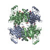 6ayuS S: Starting model for refinement C: citing same article ( |
|---|---|
| Similar structure data |
-Links
 PDBj
PDBj

- Assembly
| Deposited unit | 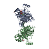
| ||||||||||||
|---|---|---|---|---|---|---|---|---|---|---|---|---|---|
| 1 | 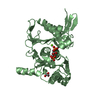
| ||||||||||||
| 2 | 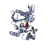
| ||||||||||||
| Unit cell |
|
-Components
| #1: Protein | Mass: 36660.430 Da / Num. of mol.: 2 Source method: isolated from a genetically manipulated source Source: (gene. exp.) Mycobacterium tuberculosis (bacteria) / Gene: glpX, MRA_1110 / Plasmid: pET-15b / Production host: #2: Chemical |   Mass: 92.094 Da / Num. of mol.: 2 / Source method: obtained synthetically / Formula: C3H8O3 Mass: 92.094 Da / Num. of mol.: 2 / Source method: obtained synthetically / Formula: C3H8O3#3: Sugar |   Type: D-saccharide, beta linking / Mass: 340.116 Da / Num. of mol.: 2 / Source method: obtained synthetically / Formula: C6H14O12P2 / Feature type: SUBJECT OF INVESTIGATION Type: D-saccharide, beta linking / Mass: 340.116 Da / Num. of mol.: 2 / Source method: obtained synthetically / Formula: C6H14O12P2 / Feature type: SUBJECT OF INVESTIGATION#4: Chemical |   Mass: 24.305 Da / Num. of mol.: 2 / Source method: obtained synthetically / Formula: Mg Mass: 24.305 Da / Num. of mol.: 2 / Source method: obtained synthetically / Formula: Mg#5: Water | ChemComp-HOH / |  Mass: 18.015 Da / Num. of mol.: 45 / Source method: isolated from a natural source / Formula: H2O Mass: 18.015 Da / Num. of mol.: 45 / Source method: isolated from a natural source / Formula: H2OHas ligand of interest | Y | |
|---|
-Experimental details
-Experiment
| Experiment | Method: X-RAY DIFFRACTION / Number of used crystals: 1 |
|---|
- Sample preparation
| Crystal | Density Matthews: 4.4 Å3/Da / Density % sol: 72.04 % / Description: hexagonal bipyramids |
|---|---|
| Crystal grow | Temperature: 291 K / Method: vapor diffusion, hanging drop / pH: 7.7 / Details: 2.9 M Sodium Malonate pH 4.0 |
-Data collection
| Diffraction | Mean temperature: 100 K / Serial crystal experiment: N |
|---|---|
| Diffraction source | Source: SYNCHROTRON / Site: APS  / Beamline: 22-ID / Wavelength: 1 Å / Beamline: 22-ID / Wavelength: 1 Å |
| Detector | Type: MAR CCD 165 mm / Detector: CCD / Date: Jul 15, 2011 |
| Radiation | Protocol: SINGLE WAVELENGTH / Monochromatic (M) / Laue (L): M / Scattering type: x-ray |
| Radiation wavelength | Wavelength: 1 Å / Relative weight: 1 |
| Reflection | Resolution: 3.71→20 Å / Num. obs: 14655 / % possible obs: 98.64 % / Redundancy: 18.4 % / Biso Wilson estimate: 88.91 Å2 / Rmerge(I) obs: 0.2 / Rrim(I) all: 0.14 / Rsym value: 0.2 / Net I/σ(I): 11 |
| Reflection shell | Resolution: 3.71→3.84 Å / Redundancy: 10.8 % / Rmerge(I) obs: 0.678 / Mean I/σ(I) obs: 1.6 / Num. unique obs: 656 / Rrim(I) all: 0.427 / Rsym value: 0.678 / % possible all: 89.76 |
- Processing
| Software |
| ||||||||||||||||||||||||||||||||||||||||||
|---|---|---|---|---|---|---|---|---|---|---|---|---|---|---|---|---|---|---|---|---|---|---|---|---|---|---|---|---|---|---|---|---|---|---|---|---|---|---|---|---|---|---|---|
| Refinement | Method to determine structure: MOLECULAR REPLACEMENT Starting model: 6ayu Resolution: 3.71→19.99 Å / SU ML: 0.3546 / Cross valid method: FREE R-VALUE / σ(F): 1.37 / Phase error: 26.368 Stereochemistry target values: GeoStd + Monomer Library + CDL v1.2
| ||||||||||||||||||||||||||||||||||||||||||
| Solvent computation | Shrinkage radii: 0.9 Å / VDW probe radii: 1.11 Å / Solvent model: FLAT BULK SOLVENT MODEL | ||||||||||||||||||||||||||||||||||||||||||
| Displacement parameters | Biso mean: 91.19 Å2 | ||||||||||||||||||||||||||||||||||||||||||
| Refinement step | Cycle: LAST / Resolution: 3.71→19.99 Å
| ||||||||||||||||||||||||||||||||||||||||||
| Refine LS restraints |
| ||||||||||||||||||||||||||||||||||||||||||
| LS refinement shell |
|
