 Movie
Movie Controller

[English] 日本語
 Yorodumi
- PDB-7txg: Structure of the Class II Fructose-1,6-Bisphosphatase from Franci... -
+ Open data

- Basic information
| Entry | Database: PDB / ID: 7txg | ||||||
|---|---|---|---|---|---|---|---|
| Title | Structure of the Class II Fructose-1,6-Bisphosphatase from Francisella tularensis with native Mn++ divalent cation and partially occupied product F6P | ||||||
 Components Components | Fructose-1,6-bisphosphatase | ||||||
 Keywords Keywords | HYDROLASE / class II FBPases / fructose-1 / 6-bisphosphatase / Francisella tularensis / Mycobacterium tuberculosis / gluconeogenesis / antibiotic targets | ||||||
| Function / homology |  Function and homology information Function and homology informationglycerol metabolic process / fructose 1,6-bisphosphate 1-phosphatase activity / fructose 1,6-bisphosphate metabolic process / gluconeogenesis / manganese ion binding / cytosol Similarity search - Function | ||||||
| Biological species |  Francisella tularensis (bacteria) Francisella tularensis (bacteria) | ||||||
| Method |  X-RAY DIFFRACTION / SYNCHROTRON / MOLECULAR REPLACEMENT / Resolution: 1.9 Å X-RAY DIFFRACTION / SYNCHROTRON / MOLECULAR REPLACEMENT / Resolution: 1.9 Å | ||||||
 Authors Authors | Abad-Zapatero, C. / Selezneva, A.I. / Harding, L.N.M. / Movahedzadeh, F. | ||||||
| Funding support | 1items
| ||||||
 Citation Citation | Journal: Plos One / Year: 2023 Title: New structures of Class II Fructose-1,6-Bisphosphatase from Francisella tularensis provide a framework for a novel catalytic mechanism for the entire class. Authors: Selezneva, A.I. / Harding, L.N.M. / Gutka, H.J. / Movahedzadeh, F. / Abad-Zapatero, C. | ||||||
| History |
|
- Structure visualization
| Structure viewer | Molecule: MolmilJmol/JSmol |
|---|
- Downloads & links
-Download
| PDBx/mmCIF format | 7txg.cif.gz | 295.1 KB | Display | PDBx/mmCIF format |
|---|---|---|---|---|
| PDB format | pdb7txg.ent.gz | 214 KB | Display | PDB format |
| PDBx/mmJSON format | 7txg.json.gz | Tree view | PDBx/mmJSON format | |
| Others |  Other downloads Other downloads |
-Validation report
| Arichive directory | https://data.pdbj.org/pub/pdb/validation_reports/tx/7txgftp://data.pdbj.org/pub/pdb/validation_reports/tx/7txg | HTTPS FTP |
|---|
-Related structure data
| Related structure data | 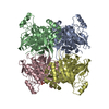 7txaC 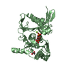 7txbC 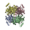 8g5wC 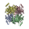 8g5xC 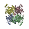 7js3S S: Starting model for refinement C: citing same article ( |
|---|---|
| Similar structure data |
-Links
 PDBj
PDBj- Assembly
| Deposited unit | 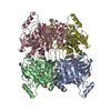
| ||||||||||||
|---|---|---|---|---|---|---|---|---|---|---|---|---|---|
| 1 |
| ||||||||||||
| Unit cell |
|
-Components
| #1: Protein | Mass: 37028.062 Da / Num. of mol.: 4 Source method: isolated from a genetically manipulated source Source: (gene. exp.) Francisella tularensis (bacteria) / Gene: glpX, FNC33_05530, FNC42_07040 / Plasmid: pET-15b / Production host: #2: Chemical | ChemComp-GOL /   Mass: 92.094 Da / Num. of mol.: 11 / Source method: obtained synthetically / Formula: C3H8O3 / Feature type: SUBJECT OF INVESTIGATION Mass: 92.094 Da / Num. of mol.: 11 / Source method: obtained synthetically / Formula: C3H8O3 / Feature type: SUBJECT OF INVESTIGATION#3: Chemical | ChemComp-MN /   Mass: 54.938 Da / Num. of mol.: 4 / Source method: obtained synthetically / Formula: Mn / Feature type: SUBJECT OF INVESTIGATION Mass: 54.938 Da / Num. of mol.: 4 / Source method: obtained synthetically / Formula: Mn / Feature type: SUBJECT OF INVESTIGATION#4: Chemical |   Mass: 94.971 Da / Num. of mol.: 3 / Source method: obtained synthetically / Formula: PO4 / Feature type: SUBJECT OF INVESTIGATION Mass: 94.971 Da / Num. of mol.: 3 / Source method: obtained synthetically / Formula: PO4 / Feature type: SUBJECT OF INVESTIGATION#5: Water | ChemComp-HOH / |  Mass: 18.015 Da / Num. of mol.: 688 / Source method: isolated from a natural source / Formula: H2O Mass: 18.015 Da / Num. of mol.: 688 / Source method: isolated from a natural source / Formula: H2OHas ligand of interest | Y | |
|---|
-Experimental details
-Experiment
| Experiment | Method: X-RAY DIFFRACTION / Number of used crystals: 1 |
|---|
- Sample preparation
| Crystal | Density Matthews: 2.34 Å3/Da / Density % sol: 47.46 % |
|---|---|
| Crystal grow | Temperature: 291 K / Method: vapor diffusion, hanging drop / pH: 8 Details: 8% Tacsimate pH 6.0 and 20% Polyethylene glycol 3350 |
-Data collection
| Diffraction | Mean temperature: 100 K / Serial crystal experiment: N |
|---|---|
| Diffraction source | Source: SYNCHROTRON / Site: APS  / Beamline: 21-ID-D / Wavelength: 1.0332 Å / Beamline: 21-ID-D / Wavelength: 1.0332 Å |
| Detector | Type: DECTRIS EIGER X 16M / Detector: PIXEL / Date: Mar 24, 2021 |
| Radiation | Protocol: SINGLE WAVELENGTH / Monochromatic (M) / Laue (L): M / Scattering type: x-ray |
| Radiation wavelength | Wavelength: 1.0332 Å / Relative weight: 1 |
| Reflection | Resolution: 1.9→40 Å / Num. obs: 85208 / % possible obs: 85.87 % / Observed criterion σ(F): 1 / Observed criterion σ(I): 1 / Redundancy: 1.8 % / Biso Wilson estimate: 27.34 Å2 / CC1/2: 0.996 / Rmerge(I) obs: 0.054 / Rpim(I) all: 0.054 / Rrim(I) all: 0.077 / Rsym value: 0.054 / Net I/σ(I): 25.27 |
| Reflection shell | Resolution: 1.9→1.97 Å / Redundancy: 1.8 % / Mean I/σ(I) obs: 4.6 / Num. unique obs: 8144 / CC1/2: 0.94 / Rpim(I) all: 0.15 / Rrim(I) all: 0.208 / % possible all: 82.5 |
- Processing
| Software |
| |||||||||||||||||||||||||||||||||||||||||||||||||||||||||||||||||||||||||||||||||||||||||||||||||||||||||||||||||||||||||||||||||||||||||||||||||||||||||||||||||||||||||||||||||||||||||||||||||||||||||||||||||||||||||
|---|---|---|---|---|---|---|---|---|---|---|---|---|---|---|---|---|---|---|---|---|---|---|---|---|---|---|---|---|---|---|---|---|---|---|---|---|---|---|---|---|---|---|---|---|---|---|---|---|---|---|---|---|---|---|---|---|---|---|---|---|---|---|---|---|---|---|---|---|---|---|---|---|---|---|---|---|---|---|---|---|---|---|---|---|---|---|---|---|---|---|---|---|---|---|---|---|---|---|---|---|---|---|---|---|---|---|---|---|---|---|---|---|---|---|---|---|---|---|---|---|---|---|---|---|---|---|---|---|---|---|---|---|---|---|---|---|---|---|---|---|---|---|---|---|---|---|---|---|---|---|---|---|---|---|---|---|---|---|---|---|---|---|---|---|---|---|---|---|---|---|---|---|---|---|---|---|---|---|---|---|---|---|---|---|---|---|---|---|---|---|---|---|---|---|---|---|---|---|---|---|---|---|---|---|---|---|---|---|---|---|---|---|---|---|---|---|---|---|
| Refinement | Method to determine structure: MOLECULAR REPLACEMENT Starting model: 7js3 Resolution: 1.9→19.94 Å / SU ML: 0.2047 / Cross valid method: FREE R-VALUE / σ(F): 2.03 / Phase error: 23.2677 Stereochemistry target values: GeoStd + Monomer Library + CDL v1.2
| |||||||||||||||||||||||||||||||||||||||||||||||||||||||||||||||||||||||||||||||||||||||||||||||||||||||||||||||||||||||||||||||||||||||||||||||||||||||||||||||||||||||||||||||||||||||||||||||||||||||||||||||||||||||||
| Solvent computation | Shrinkage radii: 0.9 Å / VDW probe radii: 1.11 Å / Solvent model: FLAT BULK SOLVENT MODEL | |||||||||||||||||||||||||||||||||||||||||||||||||||||||||||||||||||||||||||||||||||||||||||||||||||||||||||||||||||||||||||||||||||||||||||||||||||||||||||||||||||||||||||||||||||||||||||||||||||||||||||||||||||||||||
| Displacement parameters | Biso mean: 39.71 Å2 | |||||||||||||||||||||||||||||||||||||||||||||||||||||||||||||||||||||||||||||||||||||||||||||||||||||||||||||||||||||||||||||||||||||||||||||||||||||||||||||||||||||||||||||||||||||||||||||||||||||||||||||||||||||||||
| Refinement step | Cycle: LAST / Resolution: 1.9→19.94 Å
| |||||||||||||||||||||||||||||||||||||||||||||||||||||||||||||||||||||||||||||||||||||||||||||||||||||||||||||||||||||||||||||||||||||||||||||||||||||||||||||||||||||||||||||||||||||||||||||||||||||||||||||||||||||||||
| Refine LS restraints |
| |||||||||||||||||||||||||||||||||||||||||||||||||||||||||||||||||||||||||||||||||||||||||||||||||||||||||||||||||||||||||||||||||||||||||||||||||||||||||||||||||||||||||||||||||||||||||||||||||||||||||||||||||||||||||
| LS refinement shell |
|
