 Movie
Movie Controller

[English] 日本語
 Yorodumi
- PDB-7tt9: Crystal structure of Shewanella benthica Group 1 truncated hemogl... -
+ Open data

- Basic information
| Entry | Database: PDB / ID: 7tt9 | ||||||
|---|---|---|---|---|---|---|---|
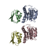
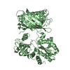
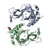
| Title | Crystal structure of Shewanella benthica Group 1 truncated hemoglobin C51S C71S Y34F variant | ||||||
 Components Components | Group 1 truncated hemoglobin | ||||||
 Keywords Keywords | HEME BINDING PROTEIN / globin / 2/2 hemoglobin / tetramer / piezophilic | ||||||
| Function / homology |  Function and homology information Function and homology information | ||||||
| Biological species |  Shewanella benthica KT99 (bacteria) Shewanella benthica KT99 (bacteria) | ||||||
| Method |  X-RAY DIFFRACTION / SAD / Resolution: 2 Å X-RAY DIFFRACTION / SAD / Resolution: 2 Å | ||||||
 Authors Authors | Martinez, J.E. / Liu, K. / Siegler, M.A. / Schlessman, J.L. / Lecomte, J.T.J. | ||||||
| Funding support |  United States, 1items United States, 1items
| ||||||
 Citation Citation | Journal: J.Biol.Chem. / Year: 2025 Title: Extremophilic hemoglobins: The structure of Shewanella benthica truncated hemoglobin N. Authors: Martinez Grundman, J.E. / Schultz, T.D. / Schlessman, J.L. / Johnson, E.A. / Gillilan, R.E. / Lecomte, J.T.J. | ||||||
| History |
|
- Structure visualization
| Structure viewer | Molecule: MolmilJmol/JSmol |
|---|
- Downloads & links
-Download
| PDBx/mmCIF format | 7tt9.cif.gz | 142.8 KB | Display | PDBx/mmCIF format |
|---|---|---|---|---|
| PDB format | pdb7tt9.ent.gz | 90.3 KB | Display | PDB format |
| PDBx/mmJSON format | 7tt9.json.gz | Tree view | PDBx/mmJSON format | |
| Others |  Other downloads Other downloads |
-Validation report
| Arichive directory | https://data.pdbj.org/pub/pdb/validation_reports/tt/7tt9ftp://data.pdbj.org/pub/pdb/validation_reports/tt/7tt9 | HTTPS FTP |
|---|
-Related structure data
| Related structure data |  8tlsC  8uzuC  8vijC  3aq7S S: Starting model for refinement C: citing same article ( |
|---|---|
| Similar structure data |

-Links
 PDBj
PDBj


- Assembly
| Deposited unit | 
| ||||||||||||||||||||||||||||||||||||||||||||||||||||||||||||||||||||||||||||||||||||||||||||||
|---|---|---|---|---|---|---|---|---|---|---|---|---|---|---|---|---|---|---|---|---|---|---|---|---|---|---|---|---|---|---|---|---|---|---|---|---|---|---|---|---|---|---|---|---|---|---|---|---|---|---|---|---|---|---|---|---|---|---|---|---|---|---|---|---|---|---|---|---|---|---|---|---|---|---|---|---|---|---|---|---|---|---|---|---|---|---|---|---|---|---|---|---|---|---|---|
| 1 |
| ||||||||||||||||||||||||||||||||||||||||||||||||||||||||||||||||||||||||||||||||||||||||||||||
| Unit cell |
| ||||||||||||||||||||||||||||||||||||||||||||||||||||||||||||||||||||||||||||||||||||||||||||||
| Noncrystallographic symmetry (NCS) | NCS domain:
NCS domain segments:
NCS oper:
|
-Components
| #1: Protein | Mass: 12822.427 Da / Num. of mol.: 4 / Mutation: C51S, C71S, Y34F Source method: isolated from a genetically manipulated source Source: (gene. exp.) Shewanella benthica KT99 (bacteria) / Gene: KT99_16901 / Production host: #2: Chemical | ChemComp-HEM /   Mass: 616.487 Da / Num. of mol.: 4 / Source method: obtained synthetically / Formula: C34H32FeN4O4 / Feature type: SUBJECT OF INVESTIGATION Mass: 616.487 Da / Num. of mol.: 4 / Source method: obtained synthetically / Formula: C34H32FeN4O4 / Feature type: SUBJECT OF INVESTIGATION#3: Water | ChemComp-HOH / |  Mass: 18.015 Da / Num. of mol.: 533 / Source method: isolated from a natural source / Formula: H2O Mass: 18.015 Da / Num. of mol.: 533 / Source method: isolated from a natural source / Formula: H2OHas ligand of interest | Y | Has protein modification | N | |
|---|
-Experimental details
-Experiment
| Experiment | Method: X-RAY DIFFRACTION / Number of used crystals: 1 |
|---|
- Sample preparation
| Crystal | Density Matthews: 2.06 Å3/Da / Density % sol: 40.4 % |
|---|---|
| Crystal grow | Temperature: 298 K / Method: vapor diffusion, hanging drop / pH: 6.5 / Details: 0.1 M Bis-Tris, pH 6.5, 28% w/v PEG2000 MME |
-Data collection
| Diffraction | Mean temperature: 110 K / Serial crystal experiment: N |
|---|---|
| Diffraction source | Source: SEALED TUBE / Type: Agilent SuperNova / Wavelength: 1.54 Å |
| Detector | Type: AGILENT ATLAS CCD / Detector: CCD / Date: Sep 16, 2021 |
| Radiation | Monochromator: MIrror / Protocol: SINGLE WAVELENGTH / Monochromatic (M) / Laue (L): M / Scattering type: x-ray |
| Radiation wavelength | Wavelength: 1.54 Å / Relative weight: 1 |
| Reflection | Resolution: 2→24.28 Å / Num. obs: 30655 / % possible obs: 99.98 % / Redundancy: 3.04 % / Biso Wilson estimate: 11.95 Å2 / CC1/2: 0.998 / Rmerge(I) obs: 0.043 / Rpim(I) all: 0.028 / Rrim(I) all: 0.051 / Net I/σ(I): 20.2 |
| Reflection shell | Resolution: 2→2.07 Å / Redundancy: 2.29 % / Rmerge(I) obs: 0.113 / Mean I/σ(I) obs: 5.8 / Num. unique obs: 5684 / CC1/2: 0.981 / Rpim(I) all: 0.091 / Rrim(I) all: 0.146 / % possible all: 99.84 |
- Processing
| Software |
| |||||||||||||||||||||||||||||||||||||||||||||||||||||||||||||||||||||||||||||||||||||||||||||||||||||||||
|---|---|---|---|---|---|---|---|---|---|---|---|---|---|---|---|---|---|---|---|---|---|---|---|---|---|---|---|---|---|---|---|---|---|---|---|---|---|---|---|---|---|---|---|---|---|---|---|---|---|---|---|---|---|---|---|---|---|---|---|---|---|---|---|---|---|---|---|---|---|---|---|---|---|---|---|---|---|---|---|---|---|---|---|---|---|---|---|---|---|---|---|---|---|---|---|---|---|---|---|---|---|---|---|---|---|---|
| Refinement | Method to determine structure: SAD Starting model: PDB entry 3AQ7 Resolution: 2→23.45 Å / SU ML: 0.17 / Cross valid method: FREE R-VALUE / σ(F): 0 / Phase error: 20.41 Stereochemistry target values: GeoStd + Monomer Library + CDL v1.2 Details: MR-SAD: rough MR solution combined with purely SAD-based structure factors obtained from anomalous data
| |||||||||||||||||||||||||||||||||||||||||||||||||||||||||||||||||||||||||||||||||||||||||||||||||||||||||
| Solvent computation | Shrinkage radii: 0.9 Å / VDW probe radii: 1.11 Å / Solvent model: FLAT BULK SOLVENT MODEL | |||||||||||||||||||||||||||||||||||||||||||||||||||||||||||||||||||||||||||||||||||||||||||||||||||||||||
| Displacement parameters | Biso mean: 14.58 Å2 | |||||||||||||||||||||||||||||||||||||||||||||||||||||||||||||||||||||||||||||||||||||||||||||||||||||||||
| Refinement step | Cycle: LAST / Resolution: 2→23.45 Å
| |||||||||||||||||||||||||||||||||||||||||||||||||||||||||||||||||||||||||||||||||||||||||||||||||||||||||
| Refine LS restraints |
| |||||||||||||||||||||||||||||||||||||||||||||||||||||||||||||||||||||||||||||||||||||||||||||||||||||||||
| Refine LS restraints NCS |
| |||||||||||||||||||||||||||||||||||||||||||||||||||||||||||||||||||||||||||||||||||||||||||||||||||||||||
| LS refinement shell |
|
