 Movie
Movie Controller

[English] 日本語
 Yorodumi
- PDB-8vij: Crystal structure of Shewanella benthica Group 1 truncated hemogl... -
+ Open data

- Basic information
| Entry | Database: PDB / ID: 8vij | ||||||
|---|---|---|---|---|---|---|---|
| Title | Crystal structure of Shewanella benthica Group 1 truncated hemoglobin Y34F C51S C71S variant (cyanomet) | ||||||
 Components Components | Group 1 truncated hemoglobin | ||||||
 Keywords Keywords | HEME-BINDING PROTEIN / GLOBIN / 2/2 HEMOGLOBIN / HEME BINDING PROTEIN / PSYCHROPIEZOPHILE | ||||||
| Function / homology |  Function and homology information Function and homology information | ||||||
| Biological species |  Shewanella benthica KT99 (bacteria) Shewanella benthica KT99 (bacteria) | ||||||
| Method |  X-RAY DIFFRACTION / SAD / Resolution: 1.35 Å X-RAY DIFFRACTION / SAD / Resolution: 1.35 Å | ||||||
 Authors Authors | Lecomte, J.T.J. / Schlessman, J.L. / Martinez, J.E. / Schultz, T.D. / Siegler, M.A. / DelCampo, M. / Le Magueres, P. | ||||||
| Funding support |  United States, 1items United States, 1items
| ||||||
 Citation Citation | Journal: J.Biol.Chem. / Year: 2025 Title: Extremophilic hemoglobins: The structure of Shewanella benthica truncated hemoglobin N. Authors: Martinez Grundman, J.E. / Schultz, T.D. / Schlessman, J.L. / Johnson, E.A. / Gillilan, R.E. / Lecomte, J.T.J. | ||||||
| History |
|
- Structure visualization
| Structure viewer | Molecule: MolmilJmol/JSmol |
|---|
- Downloads & links
-Download
| PDBx/mmCIF format | 8vij.cif.gz | 212 KB | Display | PDBx/mmCIF format |
|---|---|---|---|---|
| PDB format | pdb8vij.ent.gz | 170.6 KB | Display | PDB format |
| PDBx/mmJSON format | 8vij.json.gz | Tree view | PDBx/mmJSON format | |
| Others |  Other downloads Other downloads |
-Validation report
| Arichive directory | https://data.pdbj.org/pub/pdb/validation_reports/vi/8vijftp://data.pdbj.org/pub/pdb/validation_reports/vi/8vij | HTTPS FTP |
|---|
-Related structure data


-Links
 PDBj
PDBj


- Assembly
| Deposited unit | 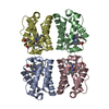
| ||||||||
|---|---|---|---|---|---|---|---|---|---|
| 1 |
| ||||||||
| Unit cell |
|
-Components
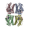
| #1: Protein | Mass: 12822.427 Da / Num. of mol.: 4 / Mutation: Y34F, C51S, C71S Source method: isolated from a genetically manipulated source Source: (gene. exp.) Shewanella benthica KT99 (bacteria) / Strain: KT99 / Gene: KT99_16901 / Production host: #2: Chemical | ChemComp-HEM /   Mass: 616.487 Da / Num. of mol.: 4 / Source method: obtained synthetically / Formula: C34H32FeN4O4 Mass: 616.487 Da / Num. of mol.: 4 / Source method: obtained synthetically / Formula: C34H32FeN4O4#3: Chemical | ChemComp-CYN /   Mass: 26.017 Da / Num. of mol.: 4 / Source method: obtained synthetically / Formula: CN Mass: 26.017 Da / Num. of mol.: 4 / Source method: obtained synthetically / Formula: CN#4: Water | ChemComp-HOH / |  Mass: 18.015 Da / Num. of mol.: 422 / Source method: isolated from a natural source / Formula: H2O Mass: 18.015 Da / Num. of mol.: 422 / Source method: isolated from a natural source / Formula: H2OHas ligand of interest | N | Has protein modification | N | |
|---|
-Experimental details
-Experiment
| Experiment | Method: X-RAY DIFFRACTION / Number of used crystals: 1 |
|---|
- Sample preparation
| Crystal | Density Matthews: 2.09 Å3/Da / Density % sol: 38.1 % |
|---|---|
| Crystal grow | Temperature: 293 K / Method: vapor diffusion, hanging drop / pH: 6.5 Details: 23% PEG MME 2000, 100 mM Bis-Tris pH 6.5, 10 mM KCN |
-Data collection
| Diffraction | Mean temperature: 100 K Ambient temp details: Temperature oscillated and increased to about 105 K first and then 110 K in the last quarter of the experiment. Serial crystal experiment: N |
|---|---|
| Diffraction source | Source: ROTATING ANODE / Type: RIGAKU MICROMAX-007 HF / Wavelength: 1.54184 Å |
| Detector | Type: RIGAKU HyPix-Arc 100 / Detector: PIXEL / Date: Jul 3, 2022 |
| Radiation | Monochromator: Rigaku VariMax DW optics (confocal multilayer) Protocol: SINGLE WAVELENGTH / Monochromatic (M) / Laue (L): M / Scattering type: x-ray |
| Radiation wavelength | Wavelength: 1.54184 Å / Relative weight: 1 |
| Reflection | Resolution: 1.35→18.97 Å / Num. obs: 97066 / % possible obs: 99 % / Redundancy: 5 % / Biso Wilson estimate: 7.83 Å2 / Rsym value: 0.081 / Net I/σ(I): 11.02 |
| Reflection shell | Resolution: 1.35→1.4 Å / Redundancy: 2.7 % / Mean I/σ(I) obs: 3.46 / Num. unique obs: 9662 / Rsym value: 0.286 / % possible all: 98.6 |
- Processing
| Software |
| |||||||||||||||||||||||||||||||||||||||||||||||||||||||||||||||||||||||||||||||||||||||||||||||||||||||||
|---|---|---|---|---|---|---|---|---|---|---|---|---|---|---|---|---|---|---|---|---|---|---|---|---|---|---|---|---|---|---|---|---|---|---|---|---|---|---|---|---|---|---|---|---|---|---|---|---|---|---|---|---|---|---|---|---|---|---|---|---|---|---|---|---|---|---|---|---|---|---|---|---|---|---|---|---|---|---|---|---|---|---|---|---|---|---|---|---|---|---|---|---|---|---|---|---|---|---|---|---|---|---|---|---|---|---|
| Refinement | Method to determine structure: SAD / Resolution: 1.35→16.97 Å / SU ML: 0.11 / Cross valid method: FREE R-VALUE / σ(F): 0.8 / Phase error: 18.45 / Stereochemistry target values: ML
| |||||||||||||||||||||||||||||||||||||||||||||||||||||||||||||||||||||||||||||||||||||||||||||||||||||||||
| Solvent computation | Shrinkage radii: 0.9 Å / VDW probe radii: 1.1 Å / Solvent model: FLAT BULK SOLVENT MODEL | |||||||||||||||||||||||||||||||||||||||||||||||||||||||||||||||||||||||||||||||||||||||||||||||||||||||||
| Refinement step | Cycle: LAST / Resolution: 1.35→16.97 Å
| |||||||||||||||||||||||||||||||||||||||||||||||||||||||||||||||||||||||||||||||||||||||||||||||||||||||||
| Refine LS restraints |
| |||||||||||||||||||||||||||||||||||||||||||||||||||||||||||||||||||||||||||||||||||||||||||||||||||||||||
| LS refinement shell |
|
