 Movie
Movie Controller

[English] 日本語
 Yorodumi
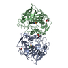
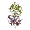
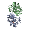
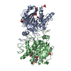
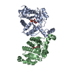
- PDB-7thi: Human Bisphosphoglycerate Mutase complexed with 2-phosphoglycolate -
+ Open data

- Basic information
| Entry | Database: PDB / ID: 7thi | ||||||
|---|---|---|---|---|---|---|---|
| Title | Human Bisphosphoglycerate Mutase complexed with 2-phosphoglycolate | ||||||
 Components Components | Bisphosphoglycerate mutase | ||||||
 Keywords Keywords | ISOMERASE / ligand / activator | ||||||
| Function / homology |  Function and homology information Function and homology informationcarbohydrate derivative catabolic process / bisphosphoglycerate mutase / bisphosphoglycerate mutase activity / phosphoglycerate mutase (2,3-diphosphoglycerate-dependent) / phosphoglycerate mutase activity / neuroinflammatory response / respiratory gaseous exchange by respiratory system / establishment of blood-brain barrier / Glycolysis / defense response to protozoan ...carbohydrate derivative catabolic process / bisphosphoglycerate mutase / bisphosphoglycerate mutase activity / phosphoglycerate mutase (2,3-diphosphoglycerate-dependent) / phosphoglycerate mutase activity / neuroinflammatory response / respiratory gaseous exchange by respiratory system / establishment of blood-brain barrier / Glycolysis / defense response to protozoan / cellular response to stress / erythrocyte development / oxygen transport / glycolytic process / carbohydrate metabolic process / hydrolase activity / extracellular exosome / cytosol Similarity search - Function | ||||||
| Biological species |  Homo sapiens (human) Homo sapiens (human) | ||||||
| Method |  X-RAY DIFFRACTION / SYNCHROTRON / MOLECULAR REPLACEMENT / Resolution: 1.33 Å X-RAY DIFFRACTION / SYNCHROTRON / MOLECULAR REPLACEMENT / Resolution: 1.33 Å | ||||||
 Authors Authors | Clark, K.L. / Kulathila, R. / Wright, K. / Isome, Y. / Sage, D. / Yang, Y. / Christodoulou, C. | ||||||
| Funding support | 1items
| ||||||
 Citation Citation | Journal: To Be Published Title: Human Bisphosphoglycerate Mutase complexed with 2-phosphoglycolate Authors: Clark, K.L. / Kulathila, R. / Wright, K. / Isome, Y. / Sage, D. / Yang, Y. / Christodoulou, C. | ||||||
| History |
|
- Structure visualization
| Structure viewer | Molecule: MolmilJmol/JSmol |
|---|
- Downloads & links
-Download
| PDBx/mmCIF format | 7thi.cif.gz | 404.9 KB | Display | PDBx/mmCIF format |
|---|---|---|---|---|
| PDB format | pdb7thi.ent.gz | 281.6 KB | Display | PDB format |
| PDBx/mmJSON format | 7thi.json.gz | Tree view | PDBx/mmJSON format | |
| Others |  Other downloads Other downloads |
-Validation report
| Summary document | 7thi_validation.pdf.gz | 1.1 MB | Display | wwPDB validaton report |
|---|---|---|---|---|
| Full document | 7thi_full_validation.pdf.gz | 1.1 MB | Display | |
| Data in XML | 7thi_validation.xml.gz | 30.3 KB | Display | |
| Data in CIF | 7thi_validation.cif.gz | 43.1 KB | Display | |
| Arichive directory | https://data.pdbj.org/pub/pdb/validation_reports/th/7thiftp://data.pdbj.org/pub/pdb/validation_reports/th/7thi | HTTPS FTP |
-Related structure data
| Related structure data |  2h4xS S: Starting model for refinement |
|---|---|
| Similar structure data |


-Links
 PDBj
PDBj


- Assembly
| Deposited unit | 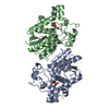
| ||||||||||||
|---|---|---|---|---|---|---|---|---|---|---|---|---|---|
| 1 |
| ||||||||||||
| Unit cell |
|
-Components
| #1: Protein | Mass: 31118.375 Da / Num. of mol.: 2 Source method: isolated from a genetically manipulated source Source: (gene. exp.) Homo sapiens (human) / Gene: BPGM / Production host:  References: UniProt: P07738, bisphosphoglycerate mutase, phosphoglycerate mutase (2,3-diphosphoglycerate-dependent) #2: Chemical | 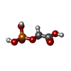  Mass: 156.031 Da / Num. of mol.: 2 / Source method: obtained synthetically / Formula: C2H5O6P / Feature type: SUBJECT OF INVESTIGATION Mass: 156.031 Da / Num. of mol.: 2 / Source method: obtained synthetically / Formula: C2H5O6P / Feature type: SUBJECT OF INVESTIGATION#3: Water | ChemComp-HOH / |  Mass: 18.015 Da / Num. of mol.: 596 / Source method: isolated from a natural source / Formula: H2O Mass: 18.015 Da / Num. of mol.: 596 / Source method: isolated from a natural source / Formula: H2OHas ligand of interest | Y | |
|---|
-Experimental details
-Experiment
| Experiment | Method: X-RAY DIFFRACTION / Number of used crystals: 1 |
|---|
- Sample preparation
| Crystal | Density Matthews: 2.23 Å3/Da / Density % sol: 44.89 % |
|---|---|
| Crystal grow | Temperature: 293 K / Method: vapor diffusion, sitting drop / pH: 8 / Details: 27% to 30% PEG 6000, bisTris propane, pH 8.0 |
-Data collection
| Diffraction | Mean temperature: 100 K / Serial crystal experiment: N | ||||||||||||||||||||||||||||||
|---|---|---|---|---|---|---|---|---|---|---|---|---|---|---|---|---|---|---|---|---|---|---|---|---|---|---|---|---|---|---|---|
| Diffraction source | Source: SYNCHROTRON / Site: SLS  / Beamline: X10SA / Wavelength: 1 Å / Beamline: X10SA / Wavelength: 1 Å | ||||||||||||||||||||||||||||||
| Detector | Type: DECTRIS PILATUS3 R 300K / Detector: PIXEL / Date: May 18, 2018 | ||||||||||||||||||||||||||||||
| Radiation | Protocol: SINGLE WAVELENGTH / Monochromatic (M) / Laue (L): M / Scattering type: x-ray | ||||||||||||||||||||||||||||||
| Radiation wavelength | Wavelength: 1 Å / Relative weight: 1 | ||||||||||||||||||||||||||||||
| Reflection | Resolution: 1.33→65.43 Å / Num. obs: 129374 / % possible obs: 100 % / Redundancy: 12.7 % / CC1/2: 0.999 / Rmerge(I) obs: 0.056 / Rpim(I) all: 0.016 / Rrim(I) all: 0.058 / Net I/σ(I): 20.2 / Num. measured all: 1638029 / Scaling rejects: 5 | ||||||||||||||||||||||||||||||
| Reflection shell | Diffraction-ID: 1
|
- Processing
| Software |
| |||||||||||||||||||||||||||||||||||||||||||||||||||||||||||||||||||||||||||||||||||||||||||||||||||||||||||||||||||||||||||||||||||||||||||||||||||||||||||||||||||||||||||||||||||||||||||||||||||||||||||||||||||||||||
|---|---|---|---|---|---|---|---|---|---|---|---|---|---|---|---|---|---|---|---|---|---|---|---|---|---|---|---|---|---|---|---|---|---|---|---|---|---|---|---|---|---|---|---|---|---|---|---|---|---|---|---|---|---|---|---|---|---|---|---|---|---|---|---|---|---|---|---|---|---|---|---|---|---|---|---|---|---|---|---|---|---|---|---|---|---|---|---|---|---|---|---|---|---|---|---|---|---|---|---|---|---|---|---|---|---|---|---|---|---|---|---|---|---|---|---|---|---|---|---|---|---|---|---|---|---|---|---|---|---|---|---|---|---|---|---|---|---|---|---|---|---|---|---|---|---|---|---|---|---|---|---|---|---|---|---|---|---|---|---|---|---|---|---|---|---|---|---|---|---|---|---|---|---|---|---|---|---|---|---|---|---|---|---|---|---|---|---|---|---|---|---|---|---|---|---|---|---|---|---|---|---|---|---|---|---|---|---|---|---|---|---|---|---|---|---|---|---|---|
| Refinement | Method to determine structure: MOLECULAR REPLACEMENT Starting model: 2H4X Resolution: 1.33→46.49 Å / SU ML: 0.1385 / Cross valid method: FREE R-VALUE / σ(F): 1.34 / Phase error: 15.1971 Stereochemistry target values: GeoStd + Monomer Library + CDL v1.2
| |||||||||||||||||||||||||||||||||||||||||||||||||||||||||||||||||||||||||||||||||||||||||||||||||||||||||||||||||||||||||||||||||||||||||||||||||||||||||||||||||||||||||||||||||||||||||||||||||||||||||||||||||||||||||
| Solvent computation | Shrinkage radii: 0.9 Å / VDW probe radii: 1.11 Å / Solvent model: FLAT BULK SOLVENT MODEL | |||||||||||||||||||||||||||||||||||||||||||||||||||||||||||||||||||||||||||||||||||||||||||||||||||||||||||||||||||||||||||||||||||||||||||||||||||||||||||||||||||||||||||||||||||||||||||||||||||||||||||||||||||||||||
| Displacement parameters | Biso mean: 25.67 Å2 | |||||||||||||||||||||||||||||||||||||||||||||||||||||||||||||||||||||||||||||||||||||||||||||||||||||||||||||||||||||||||||||||||||||||||||||||||||||||||||||||||||||||||||||||||||||||||||||||||||||||||||||||||||||||||
| Refinement step | Cycle: LAST / Resolution: 1.33→46.49 Å
| |||||||||||||||||||||||||||||||||||||||||||||||||||||||||||||||||||||||||||||||||||||||||||||||||||||||||||||||||||||||||||||||||||||||||||||||||||||||||||||||||||||||||||||||||||||||||||||||||||||||||||||||||||||||||
| Refine LS restraints |
| |||||||||||||||||||||||||||||||||||||||||||||||||||||||||||||||||||||||||||||||||||||||||||||||||||||||||||||||||||||||||||||||||||||||||||||||||||||||||||||||||||||||||||||||||||||||||||||||||||||||||||||||||||||||||
| LS refinement shell |
|
