 Movie
Movie Controller

[English] 日本語
 Yorodumi
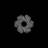
- PDB-7tdv: Crystal structure of S. aureus glutamine synthetase in Met-Sox-P/... -
+ Open data

- Basic information
| Entry | Database: PDB / ID: 7tdv | ||||||
|---|---|---|---|---|---|---|---|
| Title | Crystal structure of S. aureus glutamine synthetase in Met-Sox-P/ADP transition state complex | ||||||
 Components Components | Glutamine synthetase | ||||||
 Keywords Keywords | LIGASE / Glutamine synthetase / glutamate-ammonium ligase / GlnR / S. aureus / femC | ||||||
| Function / homology |  Function and homology information Function and homology informationglutamine synthetase / : / glutamine synthetase activity / ATP binding / metal ion binding / cytoplasm Similarity search - Function | ||||||
| Biological species |   Staphylococcus aureus (bacteria) Staphylococcus aureus (bacteria) | ||||||
| Method |  X-RAY DIFFRACTION / SYNCHROTRON / MOLECULAR REPLACEMENT / molecular replacement / Resolution: 2.92 Å X-RAY DIFFRACTION / SYNCHROTRON / MOLECULAR REPLACEMENT / molecular replacement / Resolution: 2.92 Å | ||||||
 Authors Authors | Schumacher, M.A. | ||||||
| Funding support |  United States, 1items United States, 1items
| ||||||
 Citation Citation | Journal: Nat Commun / Year: 2022 Title: Molecular dissection of the glutamine synthetase-GlnR nitrogen regulatory circuitry in Gram-positive bacteria. Authors: Brady A Travis / Jared V Peck / Raul Salinas / Brandon Dopkins / Nicholas Lent / Viet D Nguyen / Mario J Borgnia / Richard G Brennan / Maria A Schumacher / Abstract: How bacteria sense and respond to nitrogen levels are central questions in microbial physiology. In Gram-positive bacteria, nitrogen homeostasis is controlled by an operon encoding glutamine ...How bacteria sense and respond to nitrogen levels are central questions in microbial physiology. In Gram-positive bacteria, nitrogen homeostasis is controlled by an operon encoding glutamine synthetase (GS), a dodecameric machine that assimilates ammonium into glutamine, and the GlnR repressor. GlnR detects nitrogen excess indirectly by binding glutamine-feedback-inhibited-GS (FBI-GS), which activates its transcription-repression function. The molecular mechanisms behind this regulatory circuitry, however, are unknown. Here we describe biochemical and structural analyses of GS and FBI-GS-GlnR complexes from pathogenic and non-pathogenic Gram-positive bacteria. The structures show FBI-GS binds the GlnR C-terminal domain within its active-site cavity, juxtaposing two GlnR monomers to form a DNA-binding-competent GlnR dimer. The FBI-GS-GlnR interaction stabilizes the inactive GS conformation. Strikingly, this interaction also favors a remarkable dodecamer to tetradecamer transition in some GS, breaking the paradigm that all bacterial GS are dodecamers. These data thus unveil unique structural mechanisms of transcription and enzymatic regulation. | ||||||
| History |
|
- Structure visualization
| Structure viewer | Molecule: MolmilJmol/JSmol |
|---|
- Downloads & links
-Download
| PDBx/mmCIF format | 7tdv.cif.gz | 1.1 MB | Display | PDBx/mmCIF format |
|---|---|---|---|---|
| PDB format | pdb7tdv.ent.gz | Display | PDB format | |
| PDBx/mmJSON format | 7tdv.json.gz | Tree view | PDBx/mmJSON format | |
| Others |  Other downloads Other downloads |
-Validation report
| Arichive directory | https://data.pdbj.org/pub/pdb/validation_reports/td/7tdvftp://data.pdbj.org/pub/pdb/validation_reports/td/7tdv | HTTPS FTP |
|---|
-Related structure data
| Related structure data | 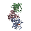 7tdpC  7teaC  7tecC 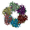 7tenC 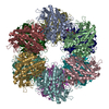 7tf6C  7tf7C 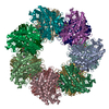 7tf9C 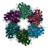 7tfaC 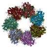 7tfbC 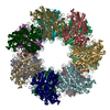 7tfcC  7tfdC  7tfeC 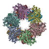 4lniS S: Starting model for refinement C: citing same article ( |
|---|---|
| Similar structure data |


-Links
 PDBj
PDBj
- Assembly
| Deposited unit | 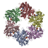
| ||||||||
|---|---|---|---|---|---|---|---|---|---|
| 1 | 
| ||||||||
| Unit cell |
|
-Components
-Protein , 1 types, 6 molecules ACDEBH
| #1: Protein | Mass: 51224.895 Da / Num. of mol.: 6 Source method: isolated from a genetically manipulated source Source: (gene. exp.) Staphylococcus aureus (bacteria)Gene: glnA, BSG37_06900, FAF32_003410, G6X35_05920, G6X37_15495, G6Y24_07770, SAMEA103891454_00902 Production host: |
|---|
-Non-polymers , 5 types, 340 molecules 
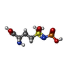


| #2: Chemical | ChemComp-ADP /  Mass: 427.201 Da / Num. of mol.: 6 / Source method: obtained synthetically / Formula: C10H15N5O10P2 / Comment: ADP, energy-carrying molecule*YM Mass: 427.201 Da / Num. of mol.: 6 / Source method: obtained synthetically / Formula: C10H15N5O10P2 / Comment: ADP, energy-carrying molecule*YM#3: Chemical | ChemComp-P3S /  Mass: 260.205 Da / Num. of mol.: 6 / Source method: obtained synthetically / Formula: C5H13N2O6PS / Feature type: SUBJECT OF INVESTIGATION Mass: 260.205 Da / Num. of mol.: 6 / Source method: obtained synthetically / Formula: C5H13N2O6PS / Feature type: SUBJECT OF INVESTIGATION#4: Chemical | ChemComp-MG /  Mass: 24.305 Da / Num. of mol.: 21 / Source method: obtained synthetically / Formula: Mg Mass: 24.305 Da / Num. of mol.: 21 / Source method: obtained synthetically / Formula: Mg#5: Chemical | ChemComp-SO4 / |  Mass: 96.063 Da / Num. of mol.: 1 / Source method: obtained synthetically / Formula: SO4 Mass: 96.063 Da / Num. of mol.: 1 / Source method: obtained synthetically / Formula: SO4#6: Water | ChemComp-HOH / | Mass: 18.015 Da / Num. of mol.: 306 / Source method: isolated from a natural source / Formula: H2O |
|---|
-Details
| Has ligand of interest | Y |
|---|
-Experimental details
-Experiment
| Experiment | Method: X-RAY DIFFRACTION / Number of used crystals: 1 |
|---|
- Sample preparation
| Crystal | Density Matthews: 2.91 Å3/Da / Density % sol: 57.73 % |
|---|---|
| Crystal grow | Temperature: 273 K / Method: vapor diffusion, hanging drop Details: 28% Peg 400, 0.2 M calcium chloride, 0.1 M HEPES 7.5 |
-Data collection
| Diffraction | Mean temperature: 100 K / Serial crystal experiment: N |
|---|---|
| Diffraction source | Source: SYNCHROTRON / Site: ALS / Beamline: 5.0.2 / Wavelength: 1.09 Å |
| Detector | Type: DECTRIS PILATUS 6M / Detector: PIXEL / Date: Dec 13, 2020 |
| Radiation | Protocol: SINGLE WAVELENGTH / Monochromatic (M) / Laue (L): M / Scattering type: x-ray |
| Radiation wavelength | Wavelength: 1.09 Å / Relative weight: 1 |
| Reflection | Resolution: 2.92→48.89 Å / Num. obs: 79309 / % possible obs: 99.9 % / Redundancy: 28 % / CC1/2: 0.999 / Rpim(I) all: 0.049 / Rsym value: 0.165 / Net I/σ(I): 19.1 |
| Reflection shell | Resolution: 2.92→3.02 Å / Mean I/σ(I) obs: 2.1 / Num. unique obs: 7783 / CC1/2: 0.744 / Rpim(I) all: 0.517 / Rsym value: 1.78 |
-Phasing
| Phasing | Method: molecular replacement |
|---|
- Processing
| Software |
| ||||||||||||||||||||||||||||||||||||||||||||||||||||||||||||||||||||||||||||||||||||
|---|---|---|---|---|---|---|---|---|---|---|---|---|---|---|---|---|---|---|---|---|---|---|---|---|---|---|---|---|---|---|---|---|---|---|---|---|---|---|---|---|---|---|---|---|---|---|---|---|---|---|---|---|---|---|---|---|---|---|---|---|---|---|---|---|---|---|---|---|---|---|---|---|---|---|---|---|---|---|---|---|---|---|---|---|---|
| Refinement | Method to determine structure: MOLECULAR REPLACEMENT Starting model: 4LNI Resolution: 2.92→48.89 Å / SU ML: 0.32 / Cross valid method: THROUGHOUT / σ(F): 1.34 / Phase error: 26.58 / Stereochemistry target values: ML
| ||||||||||||||||||||||||||||||||||||||||||||||||||||||||||||||||||||||||||||||||||||
| Solvent computation | Shrinkage radii: 0.9 Å / VDW probe radii: 1.11 Å / Solvent model: FLAT BULK SOLVENT MODEL | ||||||||||||||||||||||||||||||||||||||||||||||||||||||||||||||||||||||||||||||||||||
| Displacement parameters | Biso max: 190.94 Å2 / Biso mean: 69.5289 Å2 / Biso min: 30 Å2 | ||||||||||||||||||||||||||||||||||||||||||||||||||||||||||||||||||||||||||||||||||||
| Refinement step | Cycle: final / Resolution: 2.92→48.89 Å
| ||||||||||||||||||||||||||||||||||||||||||||||||||||||||||||||||||||||||||||||||||||
| LS refinement shell | Refine-ID: X-RAY DIFFRACTION / Rfactor Rfree error: 0 / Total num. of bins used: 13 / % reflection obs: 100 %
| ||||||||||||||||||||||||||||||||||||||||||||||||||||||||||||||||||||||||||||||||||||
| Refinement TLS params. | Method: refined / Origin x: -59.8408 Å / Origin y: 69.0059 Å / Origin z: 16.0161 Å
| ||||||||||||||||||||||||||||||||||||||||||||||||||||||||||||||||||||||||||||||||||||
| Refinement TLS group |
|
