 Movie
Movie Controller

[English] 日本語
 Yorodumi
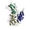
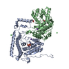
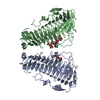
- PDB-7ta5: Crystal structure of cyanophycin synthetase 2 from Gloeothece cit... -
+ Open data

- Basic information
| Entry | Database: PDB / ID: 7ta5 | ||||||
|---|---|---|---|---|---|---|---|
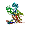
| Title | Crystal structure of cyanophycin synthetase 2 from Gloeothece citriformis | ||||||
 Components Components | Glutathione synthase | ||||||
 Keywords Keywords | LIGASE / cyanophycin / ATP-grasp / CphA | ||||||
| Function / homology |  Function and homology information Function and homology informationglutathione synthase / ribosomal S6-glutamic acid ligase activity / glutathione synthase activity / SOS response / ATP binding / metal ion binding / cytoplasm Similarity search - Function | ||||||
| Biological species |  Gloeothece citriformis (bacteria) Gloeothece citriformis (bacteria) | ||||||
| Method |  X-RAY DIFFRACTION / SYNCHROTRON / MOLECULAR REPLACEMENT / Resolution: 3 Å X-RAY DIFFRACTION / SYNCHROTRON / MOLECULAR REPLACEMENT / Resolution: 3 Å | ||||||
 Authors Authors | Sharon, I. / Schmeing, T.M. | ||||||
| Funding support |  Canada, 1items Canada, 1items
| ||||||
 Citation Citation | Journal: Acs Chem.Biol. / Year: 2022 Title: Structure and Function of the beta-Asp-Arg Polymerase Cyanophycin Synthetase 2. Authors: Sharon, I. / Grogg, M. / Hilvert, D. / Schmeing, T.M. | ||||||
| History |
|
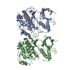
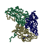
- Structure visualization
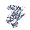
| Structure viewer | Molecule: MolmilJmol/JSmol |
|---|
- Downloads & links
-Download
| PDBx/mmCIF format | 7ta5.cif.gz | 132.4 KB | Display | PDBx/mmCIF format |
|---|---|---|---|---|
| PDB format | pdb7ta5.ent.gz | 101 KB | Display | PDB format |
| PDBx/mmJSON format | 7ta5.json.gz | Tree view | PDBx/mmJSON format | |
| Others |  Other downloads Other downloads |
-Validation report
| Summary document | 7ta5_validation.pdf.gz | 471.6 KB | Display | wwPDB validaton report |
|---|---|---|---|---|
| Full document | 7ta5_full_validation.pdf.gz | 475.5 KB | Display | |
| Data in XML | 7ta5_validation.xml.gz | 22.2 KB | Display | |
| Data in CIF | 7ta5_validation.cif.gz | 30 KB | Display | |
| Arichive directory | https://data.pdbj.org/pub/pdb/validation_reports/ta/7ta5ftp://data.pdbj.org/pub/pdb/validation_reports/ta/7ta5 | HTTPS FTP |
-Related structure data
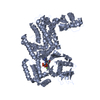
| Related structure data | 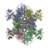 7lgjS S: Starting model for refinement |
|---|---|
| Similar structure data |

-Links
 PDBj
PDBj


- Assembly
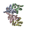
| Deposited unit | 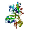
| ||||||||
|---|---|---|---|---|---|---|---|---|---|
| 1 |
| ||||||||
| Unit cell |
|
-Components
| #1: Protein | Mass: 71702.680 Da / Num. of mol.: 1 Source method: isolated from a genetically manipulated source Source: (gene. exp.) Gloeothece citriformis (bacteria) / Strain: PCC 7424 / Gene: PCC7424_0612 / Production host: | ||||||
|---|---|---|---|---|---|---|---|
| #2: Chemical | ChemComp-BCN / 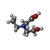  Mass: 163.172 Da / Num. of mol.: 1 / Source method: isolated from a natural source / Formula: C6H13NO4 / Comment: pH buffer*YM Mass: 163.172 Da / Num. of mol.: 1 / Source method: isolated from a natural source / Formula: C6H13NO4 / Comment: pH buffer*YM | ||||||
| #3: Chemical |   Mass: 150.087 Da / Num. of mol.: 2 / Source method: obtained synthetically / Formula: C4H6O6 Mass: 150.087 Da / Num. of mol.: 2 / Source method: obtained synthetically / Formula: C4H6O6#4: Chemical | ChemComp-TRS / |   Mass: 122.143 Da / Num. of mol.: 1 / Source method: obtained synthetically / Formula: C4H12NO3 / Comment: pH buffer*YM Mass: 122.143 Da / Num. of mol.: 1 / Source method: obtained synthetically / Formula: C4H12NO3 / Comment: pH buffer*YM#5: Water | ChemComp-HOH / |  Mass: 18.015 Da / Num. of mol.: 11 / Source method: isolated from a natural source / Formula: H2O Mass: 18.015 Da / Num. of mol.: 11 / Source method: isolated from a natural source / Formula: H2OHas ligand of interest | N | |
-Experimental details
-Experiment
| Experiment | Method: X-RAY DIFFRACTION / Number of used crystals: 1 |
|---|
- Sample preparation
| Crystal | Density Matthews: 3.77 Å3/Da / Density % sol: 67.34 % |
|---|---|
| Crystal grow | Temperature: 298 K / Method: vapor diffusion, sitting drop Details: tris-bicine pH8.5 200mM Na/K tartrate 60mM MgCl 10mM betaine 44% precip 2 4% 2,2,2-trifluoroethanol 10mM NiCl |
-Data collection
| Diffraction | Mean temperature: 100 K / Serial crystal experiment: N |
|---|---|
| Diffraction source | Source: SYNCHROTRON / Site: CLSI / Beamline: 08B1-1 / Wavelength: 1.5213 Å |
| Detector | Type: DECTRIS PILATUS3 S 6M / Detector: PIXEL / Date: Feb 28, 2021 |
| Radiation | Protocol: SINGLE WAVELENGTH / Monochromatic (M) / Laue (L): M / Scattering type: x-ray |
| Radiation wavelength | Wavelength: 1.5213 Å / Relative weight: 1 |
| Reflection | Resolution: 3→348.5 Å / Num. obs: 21675 / % possible obs: 100 % / Redundancy: 105 % / CC1/2: 0.998 / Rmerge(I) obs: 0.47 / Net I/σ(I): 18.3 |
| Reflection shell | Resolution: 3→3.18 Å / Rmerge(I) obs: 5.649 / Num. unique obs: 3378 / CC1/2: 0.451 |
- Processing
| Software |
| |||||||||||||||||||||||||||||||||||||||||||||||||||||||||||||||
|---|---|---|---|---|---|---|---|---|---|---|---|---|---|---|---|---|---|---|---|---|---|---|---|---|---|---|---|---|---|---|---|---|---|---|---|---|---|---|---|---|---|---|---|---|---|---|---|---|---|---|---|---|---|---|---|---|---|---|---|---|---|---|---|---|
| Refinement | Method to determine structure: MOLECULAR REPLACEMENT Starting model: 7LGJ Resolution: 3→116.16 Å / SU ML: 0.57 / Cross valid method: FREE R-VALUE / σ(F): 1.92 / Phase error: 31.95 / Stereochemistry target values: ML
| |||||||||||||||||||||||||||||||||||||||||||||||||||||||||||||||
| Solvent computation | Shrinkage radii: 0.9 Å / VDW probe radii: 1.11 Å / Solvent model: FLAT BULK SOLVENT MODEL | |||||||||||||||||||||||||||||||||||||||||||||||||||||||||||||||
| Refinement step | Cycle: LAST / Resolution: 3→116.16 Å
| |||||||||||||||||||||||||||||||||||||||||||||||||||||||||||||||
| Refine LS restraints |
| |||||||||||||||||||||||||||||||||||||||||||||||||||||||||||||||
| LS refinement shell |
|
