 Movie
Movie Controller


+ Open data

- Basic information
| Entry | Database: PDB / ID: 7t7n | ||||||
|---|---|---|---|---|---|---|---|
| Title | Structure of SPCC1393.13 protein from fission yeast | ||||||
 Components Components | Damage-control phosphatase SPCC1393.13 | ||||||
 Keywords Keywords | HYDROLASE / metal-dependent phosphatase / Domain of Unknown Function 89 (DUF89) | ||||||
| Function / homology |  Function and homology information Function and homology informationfructose 6-phosphate aldolase activity / fructose-1-phosphatase activity / inorganic diphosphate phosphatase activity / Hydrolases; Acting on ester bonds; Phosphoric-monoester hydrolases / intracellular phosphate ion homeostasis / cellular detoxification / phosphatase activity / DNA damage response / metal ion binding / nucleus / cytosol Similarity search - Function | ||||||
| Biological species |  | ||||||
| Method |  X-RAY DIFFRACTION / SYNCHROTRON / MOLECULAR REPLACEMENT / Resolution: 2 Å X-RAY DIFFRACTION / SYNCHROTRON / MOLECULAR REPLACEMENT / Resolution: 2 Å | ||||||
 Authors Authors | Jacewicz, A. / Sanchez, A.M. / Shuman, S. | ||||||
| Funding support |  United States, 1items United States, 1items
| ||||||
 Citation Citation | Journal: J.Biol.Chem. / Year: 2022 Title: Fission yeast Duf89 and Duf8901 are cobalt/nickel-dependent phosphatase-pyrophosphatases that act via a covalent aspartyl-phosphate intermediate. Authors: Sanchez, A.M. / Jacewicz, A. / Shuman, S. | ||||||
| History |
|
- Structure visualization
| Structure viewer | Molecule: MolmilJmol/JSmol |
|---|
- Downloads & links
-Download
| PDBx/mmCIF format | 7t7n.cif.gz | 231.9 KB | Display | PDBx/mmCIF format |
|---|---|---|---|---|
| PDB format | pdb7t7n.ent.gz | 152.6 KB | Display | PDB format |
| PDBx/mmJSON format | 7t7n.json.gz | Tree view | PDBx/mmJSON format | |
| Others |  Other downloads Other downloads |
-Validation report
| Summary document | 7t7n_validation.pdf.gz | 743 KB | Display | wwPDB validaton report |
|---|---|---|---|---|
| Full document | 7t7n_full_validation.pdf.gz | 745.1 KB | Display | |
| Data in XML | 7t7n_validation.xml.gz | 19.3 KB | Display | |
| Data in CIF | 7t7n_validation.cif.gz | 28.4 KB | Display | |
| Arichive directory | https://data.pdbj.org/pub/pdb/validation_reports/t7/7t7nftp://data.pdbj.org/pub/pdb/validation_reports/t7/7t7n | HTTPS FTP |
-Related structure data
| Related structure data |  7t7kSC  7t7oC  7u1vC  7u1xC 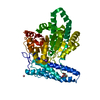 7u1yC S: Starting model for refinement C: citing same article ( |
|---|---|
| Similar structure data |
-Links
 PDBj
PDBj- Assembly
| Deposited unit | 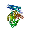
| ||||||||||||
|---|---|---|---|---|---|---|---|---|---|---|---|---|---|
| 1 |
| ||||||||||||
| Unit cell |
|
-Components
| #1: Protein | Mass: 50261.176 Da / Num. of mol.: 1 Source method: isolated from a genetically manipulated source Details: Ser 0 remains after tag removal. Three first residues from N-terminus (SMG) are disordered , therefore they were not modeled. Source: (gene. exp.) Gene: SPCC1393.13 / Production host:  References: UniProt: O94725, Hydrolases; Acting on ester bonds; Phosphoric-monoester hydrolases |
|---|---|
| #2: Chemical | ChemComp-PG5 /   Mass: 178.226 Da / Num. of mol.: 1 / Source method: obtained synthetically / Formula: C8H18O4 Mass: 178.226 Da / Num. of mol.: 1 / Source method: obtained synthetically / Formula: C8H18O4 |
| #3: Chemical | ChemComp-PO4 /   Mass: 94.971 Da / Num. of mol.: 1 / Source method: obtained synthetically / Formula: PO4 / Feature type: SUBJECT OF INVESTIGATION Mass: 94.971 Da / Num. of mol.: 1 / Source method: obtained synthetically / Formula: PO4 / Feature type: SUBJECT OF INVESTIGATION |
| #4: Water | ChemComp-HOH /  Mass: 18.015 Da / Num. of mol.: 246 / Source method: isolated from a natural source / Formula: H2O Mass: 18.015 Da / Num. of mol.: 246 / Source method: isolated from a natural source / Formula: H2O |
| Has ligand of interest | Y |
-Experimental details
-Experiment
| Experiment | Method: X-RAY DIFFRACTION / Number of used crystals: 1 |
|---|
- Sample preparation
| Crystal | Density Matthews: 2.59 Å3/Da / Density % sol: 52.55 % |
|---|---|
| Crystal grow | Temperature: 295 K / Method: vapor diffusion, sitting drop Details: 0.24 mM SPCC1393.13 protein in a buffer containing 5 mM CoCl2, 10 mM sodium pyrophosphate (pH 6.5), 18 mM Tris-HCl (pH 7.5), and 135 mM NaCl mixed with precipitant solution containing 0.1 M ...Details: 0.24 mM SPCC1393.13 protein in a buffer containing 5 mM CoCl2, 10 mM sodium pyrophosphate (pH 6.5), 18 mM Tris-HCl (pH 7.5), and 135 mM NaCl mixed with precipitant solution containing 0.1 M citric acid (pH 5.0) and 20% (w/v) PEG-6000 |
-Data collection
| Diffraction | Mean temperature: 100 K / Serial crystal experiment: N |
|---|---|
| Diffraction source | Source: SYNCHROTRON / Site: APS / Beamline: 24-ID-C / Wavelength: 1.5895 Å |
| Detector | Type: DECTRIS EIGER2 X 16M / Detector: PIXEL / Date: Oct 23, 2021 |
| Radiation | Protocol: SINGLE WAVELENGTH / Monochromatic (M) / Laue (L): M / Scattering type: x-ray |
| Radiation wavelength | Wavelength: 1.5895 Å / Relative weight: 1 |
| Reflection | Resolution: 1.99→47.36 Å / Num. obs: 31632 / % possible obs: 90.5 % / Redundancy: 6.1 % / Biso Wilson estimate: 18.4 Å2 / CC1/2: 0.995 / Rpim(I) all: 0.048 / Net I/σ(I): 12.7 |
| Reflection shell | Resolution: 1.99→2.05 Å / Mean I/σ(I) obs: 2.8 / Num. unique obs: 1004 / CC1/2: 0.797 / Rpim(I) all: 0.311 |
- Processing
| Software |
| |||||||||||||||||||||||||||||||||||||||||||||||||||||||||||||||||||||||||||||||||||||||||||||||||||||||||||||||||||||||||||||||||||||||||||||||||||||||||||||||||
|---|---|---|---|---|---|---|---|---|---|---|---|---|---|---|---|---|---|---|---|---|---|---|---|---|---|---|---|---|---|---|---|---|---|---|---|---|---|---|---|---|---|---|---|---|---|---|---|---|---|---|---|---|---|---|---|---|---|---|---|---|---|---|---|---|---|---|---|---|---|---|---|---|---|---|---|---|---|---|---|---|---|---|---|---|---|---|---|---|---|---|---|---|---|---|---|---|---|---|---|---|---|---|---|---|---|---|---|---|---|---|---|---|---|---|---|---|---|---|---|---|---|---|---|---|---|---|---|---|---|---|---|---|---|---|---|---|---|---|---|---|---|---|---|---|---|---|---|---|---|---|---|---|---|---|---|---|---|---|---|---|---|---|
| Refinement | Method to determine structure: MOLECULAR REPLACEMENT Starting model: 7T7K Resolution: 2→47.36 Å / SU ML: 0.187 / Cross valid method: FREE R-VALUE / σ(F): 1.34 / Phase error: 21.6736 Stereochemistry target values: GeoStd + Monomer Library + CDL v1.2
| |||||||||||||||||||||||||||||||||||||||||||||||||||||||||||||||||||||||||||||||||||||||||||||||||||||||||||||||||||||||||||||||||||||||||||||||||||||||||||||||||
| Solvent computation | Shrinkage radii: 0.9 Å / VDW probe radii: 1.11 Å / Solvent model: FLAT BULK SOLVENT MODEL | |||||||||||||||||||||||||||||||||||||||||||||||||||||||||||||||||||||||||||||||||||||||||||||||||||||||||||||||||||||||||||||||||||||||||||||||||||||||||||||||||
| Displacement parameters | Biso mean: 29.11 Å2 | |||||||||||||||||||||||||||||||||||||||||||||||||||||||||||||||||||||||||||||||||||||||||||||||||||||||||||||||||||||||||||||||||||||||||||||||||||||||||||||||||
| Refinement step | Cycle: LAST / Resolution: 2→47.36 Å
| |||||||||||||||||||||||||||||||||||||||||||||||||||||||||||||||||||||||||||||||||||||||||||||||||||||||||||||||||||||||||||||||||||||||||||||||||||||||||||||||||
| Refine LS restraints |
| |||||||||||||||||||||||||||||||||||||||||||||||||||||||||||||||||||||||||||||||||||||||||||||||||||||||||||||||||||||||||||||||||||||||||||||||||||||||||||||||||
| LS refinement shell |
| |||||||||||||||||||||||||||||||||||||||||||||||||||||||||||||||||||||||||||||||||||||||||||||||||||||||||||||||||||||||||||||||||||||||||||||||||||||||||||||||||
| Refinement TLS params. | Method: refined / Refine-ID: X-RAY DIFFRACTION
| |||||||||||||||||||||||||||||||||||||||||||||||||||||||||||||||||||||||||||||||||||||||||||||||||||||||||||||||||||||||||||||||||||||||||||||||||||||||||||||||||
| Refinement TLS group | Refine-ID: X-RAY DIFFRACTION / Auth asym-ID: B / Label asym-ID: A
|
