 Movie
Movie Controller


+ Open data

- Basic information
| Entry | Database: PDB / ID: 7t6b | ||||||||||||||||||||||||||||||
|---|---|---|---|---|---|---|---|---|---|---|---|---|---|---|---|---|---|---|---|---|---|---|---|---|---|---|---|---|---|---|---|
| Title | Structure of S1PR2-heterotrimeric G13 signaling complex | ||||||||||||||||||||||||||||||
 Components Components |
| ||||||||||||||||||||||||||||||
 Keywords Keywords | SIGNALING PROTEIN/IMMUNE SYSTEM / S1P / S1PR2 / GPCR / membrane protein / cryo-EM / SIGNALING PROTEIN-IMMUNE SYSTEM complex | ||||||||||||||||||||||||||||||
| Function / homology |  Function and homology information Function and homology informationD5 dopamine receptor binding / positive regulation of establishment of endothelial barrier / regulation of fibroblast migration / Rho-activating G protein-coupled receptor signaling pathway / sphingosine-1-phosphate receptor activity / Lysosphingolipid and LPA receptors / filopodium assembly / regulation of small GTPase mediated signal transduction / sphingosine-1-phosphate receptor signaling pathway / G protein-coupled peptide receptor activity ...D5 dopamine receptor binding / positive regulation of establishment of endothelial barrier / regulation of fibroblast migration / Rho-activating G protein-coupled receptor signaling pathway / sphingosine-1-phosphate receptor activity / Lysosphingolipid and LPA receptors / filopodium assembly / regulation of small GTPase mediated signal transduction / sphingosine-1-phosphate receptor signaling pathway / G protein-coupled peptide receptor activity / branching involved in blood vessel morphogenesis / NRAGE signals death through JNK / CDC42 GTPase cycle / regulation of postsynapse assembly / Rho protein signal transduction / RAC1 GTPase cycle / guanyl-nucleotide exchange factor activity / brush border membrane / platelet activation / integrin binding / G protein-coupled receptor binding / regulation of blood pressure / G protein-coupled receptor activity / G-protein beta/gamma-subunit complex binding / adenylate cyclase-modulating G protein-coupled receptor signaling pathway / Olfactory Signaling Pathway / Activation of the phototransduction cascade / G protein-coupled acetylcholine receptor signaling pathway / G beta:gamma signalling through PLC beta / Presynaptic function of Kainate receptors / Thromboxane signalling through TP receptor / Activation of G protein gated Potassium channels / Inhibition of voltage gated Ca2+ channels via Gbeta/gamma subunits / G-protein activation / melanosome / Glucagon signaling in metabolic regulation / G beta:gamma signalling through CDC42 / Prostacyclin signalling through prostacyclin receptor / Synthesis, secretion, and inactivation of Glucagon-like Peptide-1 (GLP-1) / G beta:gamma signalling through BTK / photoreceptor disc membrane / ADP signalling through P2Y purinoceptor 12 / Glucagon-type ligand receptors / Sensory perception of sweet, bitter, and umami (glutamate) taste / Adrenaline,noradrenaline inhibits insulin secretion / Vasopressin regulates renal water homeostasis via Aquaporins / regulation of cell shape / Glucagon-like Peptide-1 (GLP1) regulates insulin secretion / G alpha (z) signalling events / cellular response to catecholamine stimulus / ADP signalling through P2Y purinoceptor 1 / G beta:gamma signalling through PI3Kgamma / ADORA2B mediated anti-inflammatory cytokines production / adenylate cyclase-activating dopamine receptor signaling pathway / Cooperation of PDCL (PhLP1) and TRiC/CCT in G-protein beta folding / GPER1 signaling / cellular response to prostaglandin E stimulus / heterotrimeric G-protein complex / Inactivation, recovery and regulation of the phototransduction cascade / G alpha (12/13) signalling events / G-protein beta-subunit binding / extracellular vesicle / sensory perception of taste / presynapse / in utero embryonic development / Thrombin signalling through proteinase activated receptors (PARs) / signaling receptor complex adaptor activity / adenylate cyclase-activating G protein-coupled receptor signaling pathway / actin cytoskeleton organization / retina development in camera-type eye / fibroblast proliferation / GTPase binding / G protein activity / Ca2+ pathway / High laminar flow shear stress activates signaling by PIEZO1 and PECAM1:CDH5:KDR in endothelial cells / G alpha (i) signalling events / G alpha (s) signalling events / G alpha (q) signalling events / phospholipase C-activating G protein-coupled receptor signaling pathway / Ras protein signal transduction / cell differentiation / cell population proliferation / postsynapse / Extra-nuclear estrogen signaling / G protein-coupled receptor signaling pathway / lysosomal membrane / focal adhesion / GTPase activity / positive regulation of cell population proliferation / lipid binding / synapse / GTP binding / protein-containing complex binding / glutamatergic synapse / signal transduction / extracellular exosome / membrane / metal ion binding / nucleus / plasma membrane Similarity search - Function | ||||||||||||||||||||||||||||||
| Biological species |  Homo sapiens (human) Homo sapiens (human) | ||||||||||||||||||||||||||||||
| Method | ELECTRON MICROSCOPY / single particle reconstruction / cryo EM / Resolution: 3.19 Å | ||||||||||||||||||||||||||||||
 Authors Authors | Li, X. / Chen, H. | ||||||||||||||||||||||||||||||
| Funding support |  United States, 3items United States, 3items
| ||||||||||||||||||||||||||||||
 Citation Citation | Journal: Sci Adv / Year: 2022 Title: Structure of S1PR2-heterotrimeric G signaling complex. Authors: Hongwen Chen / Kevin Chen / Weijiao Huang / Louis M Staudt / Jason G Cyster / Xiaochun Li / Abstract: Sphingosine-1-phosphate (S1P) regulates immune cell trafficking, angiogenesis, and vascular function via its five receptors. Inherited mutations in S1P receptor 2 (S1PR2) occur in individuals with ...Sphingosine-1-phosphate (S1P) regulates immune cell trafficking, angiogenesis, and vascular function via its five receptors. Inherited mutations in S1P receptor 2 (S1PR2) occur in individuals with hearing loss, and acquired mutations in S1PR2 and G occur in a malignant lymphoma. Here, we present the cryo-electron microscopy structure of S1P-bound S1PR2 coupled to the heterotrimeric G. Interaction between S1PR2 intracellular loop 2 (ICL2) and transmembrane helix 4 confines ICL2 to engage the α5 helix of G. Transforming growth factor-α shedding assays and cell migration assays support the key roles of the residues in S1PR2-G complex assembly. The structure illuminates the mechanism of receptor disruption by disease-associated mutations. Unexpectedly, we showed that FTY720-P, an agonist of the other four S1PRs, can trigger G activation via S1PR2. S1PR2 variant can increase the activity of G considerably with FTY720-P and S1P, thus revealing a basis for S1PR drug selectivity. | ||||||||||||||||||||||||||||||
| History |
|
- Structure visualization
| Structure viewer | Molecule: MolmilJmol/JSmol |
|---|
- Downloads & links
-Download
| PDBx/mmCIF format | 7t6b.cif.gz | 216.2 KB | Display | PDBx/mmCIF format |
|---|---|---|---|---|
| PDB format | pdb7t6b.ent.gz | 163.4 KB | Display | PDB format |
| PDBx/mmJSON format | 7t6b.json.gz | Tree view | PDBx/mmJSON format | |
| Others |  Other downloads Other downloads |
-Validation report
| Arichive directory | https://data.pdbj.org/pub/pdb/validation_reports/t6/7t6bftp://data.pdbj.org/pub/pdb/validation_reports/t6/7t6b | HTTPS FTP |
|---|
-Related structure data
| Related structure data |  25712MC M: map data used to model this data C: citing same article ( |
|---|---|
| Similar structure data |
-Links
 PDBj
PDBj


- Assembly
| Deposited unit | 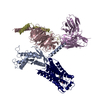
|
|---|---|
| 1 |
|
-Components
-Guanine nucleotide-binding protein ... , 3 types, 3 molecules ACD
| #1: Protein | Mass: 42255.273 Da / Num. of mol.: 1 Source method: isolated from a genetically manipulated source Source: (gene. exp.) Homo sapiens (human) / Gene: GNA13 / Cell line (production host): Sf9 / Production host:   Spodoptera frugiperda (fall armyworm) / References: UniProt: Q14344 Spodoptera frugiperda (fall armyworm) / References: UniProt: Q14344 |
|---|---|
| #2: Protein | Mass: 39970.660 Da / Num. of mol.: 1 Source method: isolated from a genetically manipulated source Source: (gene. exp.) Homo sapiens (human) / Gene: GNB1 / Cell line (production host): Sf9 / Production host: Spodoptera frugiperda (fall armyworm) / References: UniProt: P62873 |
| #3: Protein | Mass: 7861.143 Da / Num. of mol.: 1 Source method: isolated from a genetically manipulated source Source: (gene. exp.) Homo sapiens (human) / Gene: GNG2 / Production host: Spodoptera frugiperda (fall armyworm) / References: UniProt: P59768 |
-Antibody / Protein / Non-polymers , 3 types, 3 molecules ER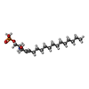
| #4: Antibody | Mass: 27784.896 Da / Num. of mol.: 1 Source method: isolated from a genetically manipulated source Source: (gene. exp.) Spodoptera frugiperda (fall armyworm) |
|---|---|
| #5: Protein | Mass: 39902.824 Da / Num. of mol.: 1 Source method: isolated from a genetically manipulated source Source: (gene. exp.) Homo sapiens (human) / Gene: S1PR2, EDG5 / Cell line (production host): HEK293S GnTI- / Production host: Homo sapiens (human) / References: UniProt: O95136 |
| #6: Chemical | ChemComp-S1P / ( Mass: 379.472 Da / Num. of mol.: 1 / Source method: obtained synthetically / Formula: C18H38NO5P / Feature type: SUBJECT OF INVESTIGATION Mass: 379.472 Da / Num. of mol.: 1 / Source method: obtained synthetically / Formula: C18H38NO5P / Feature type: SUBJECT OF INVESTIGATION |
-Details
| Has ligand of interest | Y |
|---|---|
| Has protein modification | Y |
-Experimental details
-Experiment
| Experiment | Method: ELECTRON MICROSCOPY |
|---|---|
| EM experiment | Aggregation state: PARTICLE / 3D reconstruction method: single particle reconstruction |
- Sample preparation
| Component | Name: Structure of S1PR2-heterotrimeric G13 signaling complex Type: COMPLEX / Entity ID: #1-#5 / Source: MULTIPLE SOURCES | |||||||||||||||
|---|---|---|---|---|---|---|---|---|---|---|---|---|---|---|---|---|
| Molecular weight | Experimental value: NO | |||||||||||||||
| Source (natural) |
| |||||||||||||||
| Source (recombinant) |
| |||||||||||||||
| Buffer solution | pH: 7.5 | |||||||||||||||
| Buffer component |
| |||||||||||||||
| Specimen | Embedding applied: NO / Shadowing applied: NO / Staining applied: NO / Vitrification applied: YES | |||||||||||||||
| Specimen support | Grid material: GOLD / Grid mesh size: 400 divisions/in. / Grid type: C-flat-1.2/1.3 | |||||||||||||||
| Vitrification | Cryogen name: ETHANE |
- Electron microscopy imaging
| Experimental equipment |  Model: Titan Krios / Image courtesy: FEI Company |
|---|---|
| Microscopy | Model: TFS KRIOS |
| Electron gun | Electron source:  FIELD EMISSION GUN / Accelerating voltage: 300 kV / Illumination mode: FLOOD BEAM FIELD EMISSION GUN / Accelerating voltage: 300 kV / Illumination mode: FLOOD BEAM |
| Electron lens | Mode: DIFFRACTION / Nominal defocus max: 2500 nm / Nominal defocus min: 1200 nm |
| Image recording | Electron dose: 60 e/Å2 / Film or detector model: GATAN K3 BIOQUANTUM (6k x 4k) |
- Processing
| EM software |
| ||||||||||||
|---|---|---|---|---|---|---|---|---|---|---|---|---|---|
| CTF correction | Type: NONE | ||||||||||||
| 3D reconstruction | Resolution: 3.19 Å / Resolution method: FSC 0.143 CUT-OFF / Num. of particles: 640483 / Symmetry type: POINT | ||||||||||||
| Refinement | Highest resolution: 3.19 Å |
