 Movie
Movie Controller

[English] 日本語
 Yorodumi
- PDB-7sus: Crystal structure of Apelin receptor in complex with small molecule -
+ Open data

- Basic information
| Entry | Database: PDB / ID: 7sus | ||||||
|---|---|---|---|---|---|---|---|
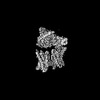
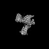
| Title | Crystal structure of Apelin receptor in complex with small molecule | ||||||
 Components Components | Apelin receptor, with Rubredoxin insertion | ||||||
 Keywords Keywords | MEMBRANE PROTEIN / GPCR / Class A GPCR / small molecule / Apelin receptor | ||||||
| Function / homology |  Function and homology information Function and homology information: / apelin receptor activity / apelin receptor signaling pathway / mechanoreceptor activity / regulation of gap junction assembly / vascular associated smooth muscle cell differentiation / atrioventricular valve development / positive regulation of G protein-coupled receptor internalization / regulation of body fluid levels / venous blood vessel development ...: / apelin receptor activity / apelin receptor signaling pathway / mechanoreceptor activity / regulation of gap junction assembly / vascular associated smooth muscle cell differentiation / atrioventricular valve development / positive regulation of G protein-coupled receptor internalization / regulation of body fluid levels / venous blood vessel development / alkane catabolic process / positive regulation of cardiac muscle hypertrophy in response to stress / endocardial cushion formation / positive regulation of blood vessel endothelial cell proliferation involved in sprouting angiogenesis / coronary vasculature development / vasculature development / G protein-coupled peptide receptor activity / adult heart development / aorta development / negative regulation of cardiac muscle hypertrophy in response to stress / ventricular septum morphogenesis / blood vessel development / heart looping / vasculogenesis / gastrulation / positive regulation of release of sequestered calcium ion into cytosol / Peptide ligand-binding receptors / G protein-coupled receptor activity / positive regulation of angiogenesis / adenylate cyclase-inhibiting G protein-coupled receptor signaling pathway / heart development / signaling receptor activity / regulation of gene expression / angiogenesis / G alpha (i) signalling events / electron transfer activity / iron ion binding / G protein-coupled receptor signaling pathway / negative regulation of gene expression / plasma membrane Similarity search - Function | ||||||
| Biological species |  Homo sapiens (human) Homo sapiens (human) Clostridium pasteurianum (bacteria) Clostridium pasteurianum (bacteria) | ||||||
| Method |  X-RAY DIFFRACTION / SYNCHROTRON / MOLECULAR REPLACEMENT / Resolution: 2.7 Å X-RAY DIFFRACTION / SYNCHROTRON / MOLECULAR REPLACEMENT / Resolution: 2.7 Å | ||||||
 Authors Authors | Xu, F. / Yue, Y. / Liu, L.E. / Han, G.W. / Hanson, M. | ||||||
| Funding support | 1items
| ||||||
 Citation Citation | Journal: Nat Struct Mol Biol / Year: 2022 Title: Structural insight into apelin receptor-G protein stoichiometry. Authors: Yang Yue / Lier Liu / Li-Jie Wu / Yiran Wu / Ling Wang / Fei Li / Junlin Liu / Gye-Won Han / Bo Chen / Xi Lin / Rebecca L Brouillette / Émile Breault / Jean-Michel Longpré / Songting Shi / ...Authors: Yang Yue / Lier Liu / Li-Jie Wu / Yiran Wu / Ling Wang / Fei Li / Junlin Liu / Gye-Won Han / Bo Chen / Xi Lin / Rebecca L Brouillette / Émile Breault / Jean-Michel Longpré / Songting Shi / Hui Lei / Philippe Sarret / Raymond C Stevens / Michael A Hanson / Fei Xu /    Abstract: The technique of cryogenic-electron microscopy (cryo-EM) has revolutionized the field of membrane protein structure and function with a focus on the dominantly observed molecular species. This report ...The technique of cryogenic-electron microscopy (cryo-EM) has revolutionized the field of membrane protein structure and function with a focus on the dominantly observed molecular species. This report describes the structural characterization of a fully active human apelin receptor (APJR) complexed with heterotrimeric G protein observed in both 2:1 and 1:1 stoichiometric ratios. We use cryo-EM single-particle analysis to determine the structural details of both species from the same sample preparation. Protein preparations, in the presence of the endogenous peptide ligand ELA or a synthetic small molecule, both demonstrate these mixed stoichiometric states. Structural differences in G protein engagement between dimeric and monomeric APJR suggest a role for the stoichiometry of G protein-coupled receptor- (GPCR-)G protein coupling on downstream signaling and receptor pharmacology. Furthermore, a small, hydrophobic dimer interface provides a starting framework for additional class A GPCR dimerization studies. Together, these findings uncover a mechanism of versatile regulation through oligomerization by which GPCRs can modulate their signaling. | ||||||
| History |
|
- Structure visualization
| Structure viewer | Molecule: MolmilJmol/JSmol |
|---|
- Downloads & links
-Download
| PDBx/mmCIF format | 7sus.cif.gz | 158.3 KB | Display | PDBx/mmCIF format |
|---|---|---|---|---|
| PDB format | pdb7sus.ent.gz | 122.2 KB | Display | PDB format |
| PDBx/mmJSON format | 7sus.json.gz | Tree view | PDBx/mmJSON format | |
| Others |  Other downloads Other downloads |
-Validation report
| Arichive directory | https://data.pdbj.org/pub/pdb/validation_reports/su/7susftp://data.pdbj.org/pub/pdb/validation_reports/su/7sus | HTTPS FTP |
|---|
-Related structure data
| Related structure data |  7w0lC  7w0mC  7w0nC  7w0oC 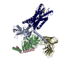 7w0pC  1iroS 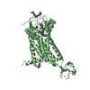 5vblS S: Starting model for refinement C: citing same article ( |
|---|---|
| Similar structure data |


-Links
 PDBj
PDBj


- Assembly
| Deposited unit | 
| ||||||||
|---|---|---|---|---|---|---|---|---|---|
| 1 |
| ||||||||
| Unit cell |
| ||||||||
| Details | The authors state that the biological oligomeric state is unknown. |
-Components
| #1: Protein | Mass: 46436.613 Da / Num. of mol.: 1 / Mutation: V117A,E174C,T177N,M217C,I250C,C325L,C326M Source method: isolated from a genetically manipulated source Source: (gene. exp.) Homo sapiens (human), (gene. exp.) Clostridium pasteurianum (bacteria)Gene: APLNR, AGTRL1, APJ / Production host:  Trichoplusia ni (cabbage looper) / References: UniProt: P35414, UniProt: P00268 Trichoplusia ni (cabbage looper) / References: UniProt: P35414, UniProt: P00268 |
|---|---|
| #2: Chemical | ChemComp-ZN /   Mass: 65.409 Da / Num. of mol.: 1 / Source method: obtained synthetically / Formula: Zn Mass: 65.409 Da / Num. of mol.: 1 / Source method: obtained synthetically / Formula: Zn |
| #3: Chemical | ChemComp-8EH / (  Mass: 525.580 Da / Num. of mol.: 1 / Source method: obtained synthetically / Formula: C24H27N7O5S / Feature type: SUBJECT OF INVESTIGATION Mass: 525.580 Da / Num. of mol.: 1 / Source method: obtained synthetically / Formula: C24H27N7O5S / Feature type: SUBJECT OF INVESTIGATION |
| #4: Chemical | ChemComp-OLC / (  Mass: 356.540 Da / Num. of mol.: 1 / Source method: obtained synthetically / Formula: C21H40O4 Mass: 356.540 Da / Num. of mol.: 1 / Source method: obtained synthetically / Formula: C21H40O4 |
| #5: Water | ChemComp-HOH /  Mass: 18.015 Da / Num. of mol.: 1 / Source method: isolated from a natural source / Formula: H2O Mass: 18.015 Da / Num. of mol.: 1 / Source method: isolated from a natural source / Formula: H2O |
| Has ligand of interest | Y |
| Has protein modification | Y |
| Sequence details | Rubredoxin from a Clostridium species is inserted in the cytoplasmic region between helices 5 and 6 |
-Experimental details
-Experiment
| Experiment | Method: X-RAY DIFFRACTION / Number of used crystals: 1 |
|---|
- Sample preparation
| Crystal | Density Matthews: 2.81 Å3/Da / Density % sol: 56.22 % |
|---|---|
| Crystal grow | Temperature: 293 K / Method: lipidic cubic phase Details: 100 mM MES pH 6.1, 26% PEG500 DME, 125 mM MgCl2, 100 mM NaCl |
-Data collection
| Diffraction | Mean temperature: 100 K / Serial crystal experiment: N |
|---|---|
| Diffraction source | Source: SYNCHROTRON / Site: SPring-8  / Beamline: BL41XU / Wavelength: 1 Å / Beamline: BL41XU / Wavelength: 1 Å |
| Detector | Type: DECTRIS PILATUS3 S 6M / Detector: PIXEL / Date: Jun 19, 2020 |
| Radiation | Protocol: SINGLE WAVELENGTH / Monochromatic (M) / Laue (L): M / Scattering type: x-ray |
| Radiation wavelength | Wavelength: 1 Å / Relative weight: 1 |
| Reflection | Resolution: 2.7→42.93 Å / Num. obs: 13301 / % possible obs: 92.3 % / Redundancy: 4.4 % / Rmerge(I) obs: 0.09 / Net I/σ(I): 5.9 |
| Reflection shell | Resolution: 2.7→2.85 Å / Rmerge(I) obs: 0.56 / Mean I/σ(I) obs: 1.3 / Num. unique obs: 1636 |
- Processing
| Software |
| ||||||||||||||||||||||||||||||||||||||||||||||||||||||||||||||||||||||||||||||||||||||||||||||||||||||||||||
|---|---|---|---|---|---|---|---|---|---|---|---|---|---|---|---|---|---|---|---|---|---|---|---|---|---|---|---|---|---|---|---|---|---|---|---|---|---|---|---|---|---|---|---|---|---|---|---|---|---|---|---|---|---|---|---|---|---|---|---|---|---|---|---|---|---|---|---|---|---|---|---|---|---|---|---|---|---|---|---|---|---|---|---|---|---|---|---|---|---|---|---|---|---|---|---|---|---|---|---|---|---|---|---|---|---|---|---|---|---|
| Refinement | Method to determine structure: MOLECULAR REPLACEMENT Starting model: 5VBL, 1IRO Resolution: 2.7→30 Å / Cor.coef. Fo:Fc: 0.919 / Cor.coef. Fo:Fc free: 0.891 / Rfactor Rfree error: 0 / SU R Cruickshank DPI: 0.755 / Cross valid method: THROUGHOUT / σ(F): 0 / SU R Blow DPI: 0.697 / SU Rfree Blow DPI: 0.335 / SU Rfree Cruickshank DPI: 0.344
| ||||||||||||||||||||||||||||||||||||||||||||||||||||||||||||||||||||||||||||||||||||||||||||||||||||||||||||
| Displacement parameters | Biso max: 274.36 Å2 / Biso mean: 129.6 Å2 / Biso min: 30 Å2
| ||||||||||||||||||||||||||||||||||||||||||||||||||||||||||||||||||||||||||||||||||||||||||||||||||||||||||||
| Refinement step | Cycle: final / Resolution: 2.7→30 Å
| ||||||||||||||||||||||||||||||||||||||||||||||||||||||||||||||||||||||||||||||||||||||||||||||||||||||||||||
| Refine LS restraints |
| ||||||||||||||||||||||||||||||||||||||||||||||||||||||||||||||||||||||||||||||||||||||||||||||||||||||||||||
| LS refinement shell | Resolution: 2.7→2.92 Å / Rfactor Rfree error: 0 / Total num. of bins used: 7
| ||||||||||||||||||||||||||||||||||||||||||||||||||||||||||||||||||||||||||||||||||||||||||||||||||||||||||||
| Refinement TLS params. | Method: refined / Refine-ID: X-RAY DIFFRACTION
| ||||||||||||||||||||||||||||||||||||||||||||||||||||||||||||||||||||||||||||||||||||||||||||||||||||||||||||
| Refinement TLS group |
|
