Movie
Movie Controller


+ Open data

- Basic information
| Entry | Database: PDB / ID: 7sj4 | |||||||||
|---|---|---|---|---|---|---|---|---|---|---|
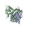
| Title | Human Trio residues 1284-1959 in complex with Rac1 | |||||||||
 Components Components |
| |||||||||
 Keywords Keywords | SIGNALING PROTEIN / guanine nucleotide exchange factor / GTPase / dbl homology / pleckstrin homology | |||||||||
| Function / homology |  Function and homology information Function and homology informationcell surface receptor protein tyrosine phosphatase signaling pathway / embryonic olfactory bulb interneuron precursor migration / anatomical structure arrangement / regulation of ERK5 cascade / angiotensin-activated signaling pathway involved in heart process / cerebral cortex GABAergic interneuron development / regulation of respiratory burst / positive regulation of ovarian follicle development / auditory receptor cell morphogenesis / cerebral cortex radially oriented cell migration ...cell surface receptor protein tyrosine phosphatase signaling pathway / embryonic olfactory bulb interneuron precursor migration / anatomical structure arrangement / regulation of ERK5 cascade / angiotensin-activated signaling pathway involved in heart process / cerebral cortex GABAergic interneuron development / regulation of respiratory burst / positive regulation of ovarian follicle development / auditory receptor cell morphogenesis / cerebral cortex radially oriented cell migration / erythrocyte enucleation / regulation of neutrophil migration / negative regulation of interleukin-23 production / Activated NTRK2 signals through CDK5 / localization within membrane / kinocilium / regulation of cell adhesion involved in heart morphogenesis / interneuron migration / ruffle assembly / NTRK2 activates RAC1 / NADPH oxidase complex / Inactivation of CDC42 and RAC1 / cochlea morphogenesis / engulfment of apoptotic cell / regulation of hydrogen peroxide metabolic process / regulation of neuron maturation / respiratory burst / WNT5:FZD7-mediated leishmania damping / SEMA3A-Plexin repulsion signaling by inhibiting Integrin adhesion / cortical cytoskeleton organization / positive regulation of skeletal muscle acetylcholine-gated channel clustering / ruffle organization / epithelial cell morphogenesis / midbrain dopaminergic neuron differentiation / positive regulation of bicellular tight junction assembly / GTP-dependent protein binding / cell projection assembly / thioesterase binding / regulation of lamellipodium assembly / negative regulation of fibroblast migration / regulation of neuron migration / regulation of stress fiber assembly / RHO GTPases activate CIT / cell-cell junction organization / Nef and signal transduction / Activation of RAC1 / motor neuron axon guidance / PCP/CE pathway / hepatocyte growth factor receptor signaling pathway / sphingosine-1-phosphate receptor signaling pathway / RHO GTPases activate KTN1 / regulation of small GTPase mediated signal transduction / regulation of nitric oxide biosynthetic process / quinolinate biosynthetic process / DCC mediated attractive signaling / MET activates RAP1 and RAC1 / Azathioprine ADME / Sema4D mediated inhibition of cell attachment and migration / hyperosmotic response / CD28 dependent Vav1 pathway / Ephrin signaling / positive regulation of neutrophil chemotaxis / superoxide anion generation / Wnt signaling pathway, planar cell polarity pathway / regulation of receptor signaling pathway via JAK-STAT / extrinsic component of membrane / lamellipodium assembly / presynaptic active zone / NRAGE signals death through JNK / positive regulation of ruffle assembly / dendrite morphogenesis / small GTPase-mediated signal transduction / Rho GDP-dissociation inhibitor binding / negative regulation of fat cell differentiation / Activation of RAC1 downstream of NMDARs / RHOJ GTPase cycle / positive regulation of Rho protein signal transduction / synaptic transmission, GABAergic / positive regulation of dendritic spine development / pericentriolar material / positive regulation of actin filament polymerization / establishment or maintenance of cell polarity / semaphorin-plexin signaling pathway / Rac protein signal transduction / RHO GTPases activate PAKs / CDC42 GTPase cycle / Sema3A PAK dependent Axon repulsion / RHOG GTPase cycle / ficolin-1-rich granule membrane / EPH-ephrin mediated repulsion of cells / positive regulation of focal adhesion assembly / regulation of postsynapse assembly / RHOA GTPase cycle / RAC3 GTPase cycle / RAC2 GTPase cycle / RHO GTPases Activate NADPH Oxidases / anatomical structure morphogenesis / regulation of synaptic vesicle endocytosis / regulation of neuronal synaptic plasticity / RHO GTPases Activate WASPs and WAVEs Similarity search - Function | |||||||||
| Biological species |  Homo sapiens (human) Homo sapiens (human) | |||||||||
| Method | ELECTRON MICROSCOPY / single particle reconstruction / cryo EM / Resolution: 2.86 Å | |||||||||
 Authors Authors | Chen, C.-L. / Ravala, S.K. / Bandekar, S.J. / Cash, J. / Tesmer, J.J.G. | |||||||||
| Funding support |  United States, 2items United States, 2items
| |||||||||
 Citation Citation | Journal: J Biol Chem / Year: 2022 Title: Structural/functional studies of Trio provide insights into its configuration and show that conserved linker elements enhance its activity for Rac1. Authors: Sumit J Bandekar / Chun-Liang Chen / Sandeep K Ravala / Jennifer N Cash / Larisa V Avramova / Mariya V Zhalnina / J Silvio Gutkind / Sheng Li / John J G Tesmer / Abstract: Trio is a large and highly conserved metazoan signaling scaffold that contains two Dbl family guanine nucleotide exchange factor (GEF) modules, TrioN and TrioC, selective for Rac and RhoA GTPases, ...Trio is a large and highly conserved metazoan signaling scaffold that contains two Dbl family guanine nucleotide exchange factor (GEF) modules, TrioN and TrioC, selective for Rac and RhoA GTPases, respectively. The GEF activities of TrioN and TrioC are implicated in several cancers, especially uveal melanoma. However, little is known about how these modules operate in the context of larger fragments of Trio. Here we show via negative stain electron microscopy that the N-terminal region of Trio is extended and could thus serve as a rigid spacer between the N-terminal putative lipid-binding domain and TrioN, whereas the C-terminal half of Trio seems globular. We found that regions C-terminal to TrioN enhance its Rac1 GEF activity and thus could play a regulatory role. We went on to characterize a minimal, well-behaved Trio fragment with enhanced activity, Trio, in complex with Rac1 using cryo-electron microscopy and hydrogen-deuterium exchange mass spectrometry and found that the region conferring enhanced activity is disordered. Deletion of two different strongly conserved motifs in this region eliminated this enhancement, suggesting that they form transient intramolecular interactions that promote GEF activity. Because Dbl family RhoGEF modules have been challenging to directly target with small molecules, characterization of accessory Trio domains such as these may provide alternate routes for the development of therapeutics that inhibit Trio activity in human cancer. | |||||||||
| History |
|
- Structure visualization
| Structure viewer | Molecule: MolmilJmol/JSmol |
|---|
- Downloads & links
-Download
| PDBx/mmCIF format | 7sj4.cif.gz | 119.8 KB | Display | PDBx/mmCIF format |
|---|---|---|---|---|
| PDB format | pdb7sj4.ent.gz | 83.9 KB | Display | PDB format |
| PDBx/mmJSON format | 7sj4.json.gz | Tree view | PDBx/mmJSON format | |
| Others |  Other downloads Other downloads |
-Validation report
| Arichive directory | https://data.pdbj.org/pub/pdb/validation_reports/sj/7sj4ftp://data.pdbj.org/pub/pdb/validation_reports/sj/7sj4 | HTTPS FTP |
|---|
-Related structure data
| Related structure data |  25153MC M: map data used to model this data C: citing same article ( |
|---|---|
| Similar structure data |
-Links
 PDBj
PDBj


- Assembly
| Deposited unit | 
|
|---|---|
| 1 |
|
-Components
| #1: Protein | Mass: 75592.969 Da / Num. of mol.: 1 Source method: isolated from a genetically manipulated source Source: (gene. exp.) Homo sapiens (human) / Gene: TRIO / Production host:  References: UniProt: O75962, non-specific serine/threonine protein kinase |
|---|---|
| #2: Protein | Mass: 21811.453 Da / Num. of mol.: 1 Source method: isolated from a genetically manipulated source Source: (gene. exp.) Homo sapiens (human) / Gene: RAC1, TC25, MIG5 / Production host: |
| #3: Water | ChemComp-HOH /  Mass: 18.015 Da / Num. of mol.: 24 / Source method: isolated from a natural source / Formula: H2O Mass: 18.015 Da / Num. of mol.: 24 / Source method: isolated from a natural source / Formula: H2O |
-Experimental details
-Experiment
| Experiment | Method: ELECTRON MICROSCOPY |
|---|---|
| EM experiment | Aggregation state: PARTICLE / 3D reconstruction method: single particle reconstruction |
- Sample preparation
| Component | Name: Human Trio residues 1284-1959 in complex with Rac1 / Type: COMPLEX / Entity ID: #1-#2 / Source: RECOMBINANT | ||||||||||||||||||||
|---|---|---|---|---|---|---|---|---|---|---|---|---|---|---|---|---|---|---|---|---|---|
| Molecular weight | Experimental value: NO | ||||||||||||||||||||
| Source (natural) | Organism: Homo sapiens (human) | ||||||||||||||||||||
| Source (recombinant) | Organism: | ||||||||||||||||||||
| Buffer solution | pH: 8 | ||||||||||||||||||||
| Buffer component |
| ||||||||||||||||||||
| Specimen | Conc.: 0.25 mg/ml / Embedding applied: NO / Shadowing applied: NO / Staining applied: NO / Vitrification applied: YES | ||||||||||||||||||||
| Specimen support | Grid material: GOLD / Grid mesh size: 300 divisions/in. / Grid type: UltrAuFoil R1.2/1.3 | ||||||||||||||||||||
| Vitrification | Instrument: FEI VITROBOT MARK IV / Cryogen name: ETHANE / Humidity: 100 % / Chamber temperature: 277 K / Details: Blot force 10 |
- Electron microscopy imaging
| Experimental equipment |  Model: Titan Krios / Image courtesy: FEI Company |
|---|---|
| Microscopy | Model: FEI TITAN KRIOS |
| Electron gun | Electron source:  FIELD EMISSION GUN / Accelerating voltage: 300 kV / Illumination mode: FLOOD BEAM FIELD EMISSION GUN / Accelerating voltage: 300 kV / Illumination mode: FLOOD BEAM |
| Electron lens | Mode: BRIGHT FIELD / Nominal magnification: 81000 X / Nominal defocus max: 3000 nm / Nominal defocus min: 1000 nm / Cs: 2.7 mm / C2 aperture diameter: 100 µm / Alignment procedure: COMA FREE |
| Specimen holder | Cryogen: NITROGEN / Specimen holder model: FEI TITAN KRIOS AUTOGRID HOLDER / Residual tilt: 0.01 mradians |
| Image recording | Average exposure time: 3.12 sec. / Electron dose: 55 e/Å2 / Detector mode: SUPER-RESOLUTION / Film or detector model: GATAN K3 BIOQUANTUM (6k x 4k) / Num. of real images: 2817 Details: 3514 movies were initially collected. After motion correction and CTF estimation, 697 micrographs were rejected due to poor CTF fitting. |
| Image scans | Width: 11520 / Height: 8184 / Movie frames/image: 40 |
- Processing
| Software | Name: PHENIX / Version: 1.20_4459: / Classification: refinement | ||||||||||||||||||||||||||||||||||||||||||||||||
|---|---|---|---|---|---|---|---|---|---|---|---|---|---|---|---|---|---|---|---|---|---|---|---|---|---|---|---|---|---|---|---|---|---|---|---|---|---|---|---|---|---|---|---|---|---|---|---|---|---|
| EM software |
| ||||||||||||||||||||||||||||||||||||||||||||||||
| CTF correction | Type: PHASE FLIPPING AND AMPLITUDE CORRECTION | ||||||||||||||||||||||||||||||||||||||||||||||||
| Symmetry | Point symmetry: C1 (asymmetric) | ||||||||||||||||||||||||||||||||||||||||||||||||
| 3D reconstruction | Resolution: 2.86 Å / Resolution method: FSC 0.143 CUT-OFF / Num. of particles: 922202 / Algorithm: BACK PROJECTION / Symmetry type: POINT | ||||||||||||||||||||||||||||||||||||||||||||||||
| Atomic model building | Protocol: RIGID BODY FIT / Space: REAL / Target criteria: correlation coefficient Details: The initial model was generated from the pdb structure (2NZ8) using the SWISS-MODEL server and rigid-body fitted using Chimera. Several runs of structure refinement were done using the coot ...Details: The initial model was generated from the pdb structure (2NZ8) using the SWISS-MODEL server and rigid-body fitted using Chimera. Several runs of structure refinement were done using the coot and phenix real-space refinement. | ||||||||||||||||||||||||||||||||||||||||||||||||
| Atomic model building | 3D fitting-ID: 1 / Accession code: 2NZ8 / Initial refinement model-ID: 1 / PDB-ID: 2NZ8 / Source name: PDB / Type: experimental model
| ||||||||||||||||||||||||||||||||||||||||||||||||
| Refine LS restraints |
|