 Movie
Movie Controller

[English] 日本語
 Yorodumi
- PDB-7sh3: Crystal Structure of a VirB8-like Protein of Type IV Secretion Sy... -
+ Open data

- Basic information
| Entry | Database: PDB / ID: 7sh3 | ||||||
|---|---|---|---|---|---|---|---|
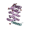
| Title | Crystal Structure of a VirB8-like Protein of Type IV Secretion System from Rickettsia typhi in Complex with a Synthetic VirB8 Miniprotein Binder | ||||||
 Components Components |
| ||||||
 Keywords Keywords | PROTEIN TRANSPORT / SSGCID / synthetic / miniprotein binder / minibinder / VirB8 / Structural Genomics / Seattle Structural Genomics Center for Infectious Disease | ||||||
| Function / homology | Bacterial virulence protein VirB8 / VirB8 protein / NTF2-like domain superfamily / membrane / VirB8-like protein of type IV secretion system Function and homology information Function and homology information | ||||||
| Biological species | synthetic construct (others) Rickettsia typhi (bacteria) Rickettsia typhi (bacteria) | ||||||
| Method |  X-RAY DIFFRACTION / SYNCHROTRON / MOLECULAR REPLACEMENT / molecular replacement / Resolution: 3 Å X-RAY DIFFRACTION / SYNCHROTRON / MOLECULAR REPLACEMENT / molecular replacement / Resolution: 3 Å | ||||||
 Authors Authors | Seattle Structural Genomics Center for Infectious Disease / Seattle Structural Genomics Center for Infectious Disease (SSGCID) | ||||||
| Funding support | 1items
| ||||||
 Citation Citation | Journal: to be published Title: Crystal Structure of a VirB8-like Protein of Type IV Secretion System from Rickettsia typhi in Complex with a Synthetic VirB8 Miniprotein Binder Authors: DeBouver, N.D. / Bera, A.K. / Lorimer, D.D. / Horanyi, P.S. / Edwards, T.E. | ||||||
| History |
|
- Structure visualization
| Structure viewer | Molecule: MolmilJmol/JSmol |
|---|
- Downloads & links
-Download
| PDBx/mmCIF format | 7sh3.cif.gz | 96 KB | Display | PDBx/mmCIF format |
|---|---|---|---|---|
| PDB format | pdb7sh3.ent.gz | 71.7 KB | Display | PDB format |
| PDBx/mmJSON format | 7sh3.json.gz | Tree view | PDBx/mmJSON format | |
| Others |  Other downloads Other downloads |
-Validation report
| Arichive directory | https://data.pdbj.org/pub/pdb/validation_reports/sh/7sh3ftp://data.pdbj.org/pub/pdb/validation_reports/sh/7sh3 | HTTPS FTP |
|---|
-Related structure data
| Related structure data | 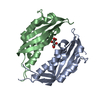 4o3vS S: Starting model for refinement |
|---|---|
| Similar structure data | |
| Other databases |


-Links
 PDBj
PDBj
- Assembly
| Deposited unit | 
| ||||||||
|---|---|---|---|---|---|---|---|---|---|
| 1 |
| ||||||||
| Unit cell |
|
-Components
| #1: Protein | Mass: 7615.714 Da / Num. of mol.: 1 Source method: isolated from a genetically manipulated source Source: (gene. exp.) synthetic construct (others) / Gene: synthetic / Plasmid: SycoA.20508.e.PG1 / Production host: |
|---|---|
| #2: Protein | Mass: 21209.270 Da / Num. of mol.: 1 Source method: isolated from a genetically manipulated source Source: (gene. exp.) Rickettsia typhi (strain ATCC VR-144 / Wilmington) (bacteria)Strain: ATCC VR-144 / Wilmington / Gene: RT0278 / Plasmid: RityA.18390.a.B2 / Production host: |
-Experimental details
-Experiment
| Experiment | Method: X-RAY DIFFRACTION / Number of used crystals: 1 |
|---|
- Sample preparation
| Crystal | Density Matthews: 2.75 Å3/Da / Density % sol: 55.32 % |
|---|---|
| Crystal grow | Temperature: 287 K / Method: vapor diffusion, sitting drop / pH: 4.6 Details: RityA.18390.a.B2 + SycoA.20508.e.PG1 [Barcode: 320527g9, PuckID: oce7-3, Cryo: 15% EG, Concentration: 9.9 mg/mL] 100mM Sodium acetate/Hydrochloric acid pH 4.6, 25% (w/v) PEG 4000, 200mM Ammonium sulfate |
-Data collection
| Diffraction | Mean temperature: 100 K / Serial crystal experiment: N | ||||||||||||||||||||||||||||||||||||||||||||||||||||||||||||||||||||||||||||||||||||||||||||||||||||||||||||||||||||||||||||||||||||||||||||||||||||||||||||||||||||||||||||||||||||||||||||||||||||||||||||||||||
|---|---|---|---|---|---|---|---|---|---|---|---|---|---|---|---|---|---|---|---|---|---|---|---|---|---|---|---|---|---|---|---|---|---|---|---|---|---|---|---|---|---|---|---|---|---|---|---|---|---|---|---|---|---|---|---|---|---|---|---|---|---|---|---|---|---|---|---|---|---|---|---|---|---|---|---|---|---|---|---|---|---|---|---|---|---|---|---|---|---|---|---|---|---|---|---|---|---|---|---|---|---|---|---|---|---|---|---|---|---|---|---|---|---|---|---|---|---|---|---|---|---|---|---|---|---|---|---|---|---|---|---|---|---|---|---|---|---|---|---|---|---|---|---|---|---|---|---|---|---|---|---|---|---|---|---|---|---|---|---|---|---|---|---|---|---|---|---|---|---|---|---|---|---|---|---|---|---|---|---|---|---|---|---|---|---|---|---|---|---|---|---|---|---|---|---|---|---|---|---|---|---|---|---|---|---|---|---|---|---|---|---|
| Diffraction source | Source: SYNCHROTRON / Site: APS  / Beamline: 21-ID-F / Wavelength: 0.9787 Å / Beamline: 21-ID-F / Wavelength: 0.9787 Å | ||||||||||||||||||||||||||||||||||||||||||||||||||||||||||||||||||||||||||||||||||||||||||||||||||||||||||||||||||||||||||||||||||||||||||||||||||||||||||||||||||||||||||||||||||||||||||||||||||||||||||||||||||
| Detector | Type: RAYONIX MX-300 / Detector: CCD / Date: Apr 15, 2021 / Details: Beryllium Lenses | ||||||||||||||||||||||||||||||||||||||||||||||||||||||||||||||||||||||||||||||||||||||||||||||||||||||||||||||||||||||||||||||||||||||||||||||||||||||||||||||||||||||||||||||||||||||||||||||||||||||||||||||||||
| Radiation | Monochromator: Diamond [111] / Protocol: SINGLE WAVELENGTH / Monochromatic (M) / Laue (L): M / Scattering type: x-ray | ||||||||||||||||||||||||||||||||||||||||||||||||||||||||||||||||||||||||||||||||||||||||||||||||||||||||||||||||||||||||||||||||||||||||||||||||||||||||||||||||||||||||||||||||||||||||||||||||||||||||||||||||||
| Radiation wavelength | Wavelength: 0.9787 Å / Relative weight: 1 | ||||||||||||||||||||||||||||||||||||||||||||||||||||||||||||||||||||||||||||||||||||||||||||||||||||||||||||||||||||||||||||||||||||||||||||||||||||||||||||||||||||||||||||||||||||||||||||||||||||||||||||||||||
| Reflection | Resolution: 3→44.69 Å / Num. obs: 6672 / % possible obs: 99.8 % / Redundancy: 5.897 % / Biso Wilson estimate: 86.87 Å2 / CC1/2: 0.999 / Rmerge(I) obs: 0.074 / Rrim(I) all: 0.082 / Χ2: 0.889 / Net I/σ(I): 13.47 / Num. measured all: 39344 | ||||||||||||||||||||||||||||||||||||||||||||||||||||||||||||||||||||||||||||||||||||||||||||||||||||||||||||||||||||||||||||||||||||||||||||||||||||||||||||||||||||||||||||||||||||||||||||||||||||||||||||||||||
| Reflection shell | Diffraction-ID: 1
|
-Phasing
| Phasing | Method: molecular replacement |
|---|
- Processing
| Software |
| |||||||||||||||||||||||||||||||||||||||||||||||||||||||||||||||||||||||||||||||||||||||||||||||||||||||||||||||||||||||||||||||||||||||||||||||||||||||||||||||||||||||||||||||
|---|---|---|---|---|---|---|---|---|---|---|---|---|---|---|---|---|---|---|---|---|---|---|---|---|---|---|---|---|---|---|---|---|---|---|---|---|---|---|---|---|---|---|---|---|---|---|---|---|---|---|---|---|---|---|---|---|---|---|---|---|---|---|---|---|---|---|---|---|---|---|---|---|---|---|---|---|---|---|---|---|---|---|---|---|---|---|---|---|---|---|---|---|---|---|---|---|---|---|---|---|---|---|---|---|---|---|---|---|---|---|---|---|---|---|---|---|---|---|---|---|---|---|---|---|---|---|---|---|---|---|---|---|---|---|---|---|---|---|---|---|---|---|---|---|---|---|---|---|---|---|---|---|---|---|---|---|---|---|---|---|---|---|---|---|---|---|---|---|---|---|---|---|---|---|---|---|
| Refinement | Method to determine structure: MOLECULAR REPLACEMENT Starting model: 4O3V Resolution: 3→44.69 Å / SU ML: 0.41 / Cross valid method: THROUGHOUT / σ(F): 1.34 / Phase error: 43.04 / Stereochemistry target values: ML
| |||||||||||||||||||||||||||||||||||||||||||||||||||||||||||||||||||||||||||||||||||||||||||||||||||||||||||||||||||||||||||||||||||||||||||||||||||||||||||||||||||||||||||||||
| Solvent computation | Shrinkage radii: 0.9 Å / VDW probe radii: 1.11 Å / Solvent model: FLAT BULK SOLVENT MODEL | |||||||||||||||||||||||||||||||||||||||||||||||||||||||||||||||||||||||||||||||||||||||||||||||||||||||||||||||||||||||||||||||||||||||||||||||||||||||||||||||||||||||||||||||
| Displacement parameters | Biso max: 218.91 Å2 / Biso mean: 129.2413 Å2 / Biso min: 91.9 Å2 | |||||||||||||||||||||||||||||||||||||||||||||||||||||||||||||||||||||||||||||||||||||||||||||||||||||||||||||||||||||||||||||||||||||||||||||||||||||||||||||||||||||||||||||||
| Refinement step | Cycle: final / Resolution: 3→44.69 Å
| |||||||||||||||||||||||||||||||||||||||||||||||||||||||||||||||||||||||||||||||||||||||||||||||||||||||||||||||||||||||||||||||||||||||||||||||||||||||||||||||||||||||||||||||
| LS refinement shell | Refine-ID: X-RAY DIFFRACTION / Rfactor Rfree error: 0 / Total num. of bins used: 5
| |||||||||||||||||||||||||||||||||||||||||||||||||||||||||||||||||||||||||||||||||||||||||||||||||||||||||||||||||||||||||||||||||||||||||||||||||||||||||||||||||||||||||||||||
| Refinement TLS params. | Method: refined / Refine-ID: X-RAY DIFFRACTION
| |||||||||||||||||||||||||||||||||||||||||||||||||||||||||||||||||||||||||||||||||||||||||||||||||||||||||||||||||||||||||||||||||||||||||||||||||||||||||||||||||||||||||||||||
| Refinement TLS group |
|
