 Movie
Movie Controller

[English] 日本語
 Yorodumi
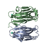
- PDB-4o3v: Crystal structure of a VirB8-like protein of type IV secretion sy... -
+ Open data

- Basic information
| Entry | Database: PDB / ID: 4o3v | ||||||
|---|---|---|---|---|---|---|---|
| Title | Crystal structure of a VirB8-like protein of type IV secretion system from Rickettsia typhi | ||||||
 Components Components | VirB8-like protein of type IV secretion system | ||||||
 Keywords Keywords | PROTEIN TRANSPORT / Structural Genomics / NIAID / National Institute of Allergy and Infectious Diseases / Seattle Structural Genomics Center for Infectious Disease / SSGCID / secretion system / TrwG / murine / typhus / virulence / pathogenesis | ||||||
| Function / homology |  Function and homology information Function and homology information | ||||||
| Biological species |  Rickettsia typhi (bacteria) Rickettsia typhi (bacteria) | ||||||
| Method |  X-RAY DIFFRACTION / SYNCHROTRON / MOLECULAR REPLACEMENT / molecular replacement / Resolution: 1.95 Å X-RAY DIFFRACTION / SYNCHROTRON / MOLECULAR REPLACEMENT / molecular replacement / Resolution: 1.95 Å | ||||||
 Authors Authors | Seattle Structural Genomics Center for Infectious Disease (SSGCID) | ||||||
 Citation Citation | Journal: MBio / Year: 2015 Title: Structural Insight into How Bacteria Prevent Interference between Multiple Divergent Type IV Secretion Systems. Authors: Gillespie, J.J. / Phan, I.Q. / Scheib, H. / Subramanian, S. / Edwards, T.E. / Lehman, S.S. / Piitulainen, H. / Rahman, M.S. / Rennoll-Bankert, K.E. / Staker, B.L. / Taira, S. / Stacy, R. / ...Authors: Gillespie, J.J. / Phan, I.Q. / Scheib, H. / Subramanian, S. / Edwards, T.E. / Lehman, S.S. / Piitulainen, H. / Rahman, M.S. / Rennoll-Bankert, K.E. / Staker, B.L. / Taira, S. / Stacy, R. / Myler, P.J. / Azad, A.F. / Pulliainen, A.T. #1: Journal: Pathog Dis / Year: 2016 Title: The Rickettsia type IV secretion system: unrealized complexity mired by gene family expansion. Authors: Gillespie, J.J. / Phan, I.Q. / Driscoll, T.P. / Guillotte, M.L. / Lehman, S.S. / Rennoll-Bankert, K.E. / Subramanian, S. / Beier-Sexton, M. / Myler, P.J. / Rahman, M.S. / Azad, A.F. | ||||||
| History |
|
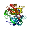
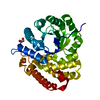
- Structure visualization
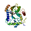
| Structure viewer | Molecule: MolmilJmol/JSmol |
|---|
- Downloads & links
-Download
| PDBx/mmCIF format | 4o3v.cif.gz | 135.5 KB | Display | PDBx/mmCIF format |
|---|---|---|---|---|
| PDB format | pdb4o3v.ent.gz | 106.3 KB | Display | PDB format |
| PDBx/mmJSON format | 4o3v.json.gz | Tree view | PDBx/mmJSON format | |
| Others |  Other downloads Other downloads |
-Validation report
| Arichive directory | https://data.pdbj.org/pub/pdb/validation_reports/o3/4o3vftp://data.pdbj.org/pub/pdb/validation_reports/o3/4o3v | HTTPS FTP |
|---|
-Related structure data
| Related structure data |  4jf8C 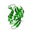 4kz1C  4lsoC  4meiC  4nhfC C: citing same article ( |
|---|---|
| Similar structure data | |
| Other databases |


-Links
 PDBj
PDBj
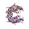
- Assembly
| Deposited unit | 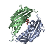
| ||||||||
|---|---|---|---|---|---|---|---|---|---|
| 1 |
| ||||||||
| Unit cell |
|
-Components
| #1: Protein | Mass: 21209.270 Da / Num. of mol.: 2 Source method: isolated from a genetically manipulated source Source: (gene. exp.) Rickettsia typhi (bacteria) / Strain: VR-144 / Willmington / Gene: RT0278 / Production host: #2: Chemical | ChemComp-SRT / |   Mass: 150.087 Da / Num. of mol.: 1 / Source method: obtained synthetically / Formula: C4H6O6 Mass: 150.087 Da / Num. of mol.: 1 / Source method: obtained synthetically / Formula: C4H6O6#3: Water | ChemComp-HOH / |  Mass: 18.015 Da / Num. of mol.: 318 / Source method: isolated from a natural source / Formula: H2O Mass: 18.015 Da / Num. of mol.: 318 / Source method: isolated from a natural source / Formula: H2O |
|---|
-Experimental details
-Experiment
| Experiment | Method: X-RAY DIFFRACTION / Number of used crystals: 1 |
|---|
- Sample preparation
| Crystal | Density Matthews: 3.42 Å3/Da / Density % sol: 64.04 % |
|---|---|
| Crystal grow | Temperature: 289 K / Method: vapor diffusion, sitting drop / pH: 7 Details: RityA.18390.a.B2 PS01633 at 11.2 mg/mL against CSHT A2, 0.4 M potassium sodium tartrate, cryo-protected with Al's oil, crystal grew over 7 months, crystal tracking ID 250519a2, unique puck ...Details: RityA.18390.a.B2 PS01633 at 11.2 mg/mL against CSHT A2, 0.4 M potassium sodium tartrate, cryo-protected with Al's oil, crystal grew over 7 months, crystal tracking ID 250519a2, unique puck ID zuk7-16, VAPOR DIFFUSION, SITTING DROP, temperature 289K |
-Data collection
| Diffraction | Mean temperature: 100 K | |||||||||||||||||||||||||||||||||||||||||||||||||||||||||||||||||||||||||||||||||||||||||||||||||||||||||||||||||||||||||||||||||||||||||||||||||||
|---|---|---|---|---|---|---|---|---|---|---|---|---|---|---|---|---|---|---|---|---|---|---|---|---|---|---|---|---|---|---|---|---|---|---|---|---|---|---|---|---|---|---|---|---|---|---|---|---|---|---|---|---|---|---|---|---|---|---|---|---|---|---|---|---|---|---|---|---|---|---|---|---|---|---|---|---|---|---|---|---|---|---|---|---|---|---|---|---|---|---|---|---|---|---|---|---|---|---|---|---|---|---|---|---|---|---|---|---|---|---|---|---|---|---|---|---|---|---|---|---|---|---|---|---|---|---|---|---|---|---|---|---|---|---|---|---|---|---|---|---|---|---|---|---|---|---|---|---|
| Diffraction source | Source: SYNCHROTRON / Site: ALS  / Beamline: 5.0.2 / Wavelength: 1 Å / Beamline: 5.0.2 / Wavelength: 1 Å | |||||||||||||||||||||||||||||||||||||||||||||||||||||||||||||||||||||||||||||||||||||||||||||||||||||||||||||||||||||||||||||||||||||||||||||||||||
| Detector | Type: ADSC QUANTUM 315r / Detector: CCD / Date: Dec 4, 2013 | |||||||||||||||||||||||||||||||||||||||||||||||||||||||||||||||||||||||||||||||||||||||||||||||||||||||||||||||||||||||||||||||||||||||||||||||||||
| Radiation | Protocol: SINGLE WAVELENGTH / Monochromatic (M) / Laue (L): M / Scattering type: x-ray | |||||||||||||||||||||||||||||||||||||||||||||||||||||||||||||||||||||||||||||||||||||||||||||||||||||||||||||||||||||||||||||||||||||||||||||||||||
| Radiation wavelength | Wavelength: 1 Å / Relative weight: 1 | |||||||||||||||||||||||||||||||||||||||||||||||||||||||||||||||||||||||||||||||||||||||||||||||||||||||||||||||||||||||||||||||||||||||||||||||||||
| Reflection | Resolution: 1.95→50 Å / Num. obs: 43393 / % possible obs: 100 % / Observed criterion σ(I): -3 / Redundancy: 9.6 % / Biso Wilson estimate: 30.753 Å2 / Rmerge(I) obs: 0.065 / Net I/σ(I): 24.26 | |||||||||||||||||||||||||||||||||||||||||||||||||||||||||||||||||||||||||||||||||||||||||||||||||||||||||||||||||||||||||||||||||||||||||||||||||||
| Reflection shell | Diffraction-ID: 1
|
-Phasing
| Phasing | Method: molecular replacement |
|---|---|
| Phasing MR | Model details: Phaser MODE: MR_AUTO |
- Processing
| Software |
| |||||||||||||||||||||||||||||||||||||||||||||||||||||||||||||||||||||||||||
|---|---|---|---|---|---|---|---|---|---|---|---|---|---|---|---|---|---|---|---|---|---|---|---|---|---|---|---|---|---|---|---|---|---|---|---|---|---|---|---|---|---|---|---|---|---|---|---|---|---|---|---|---|---|---|---|---|---|---|---|---|---|---|---|---|---|---|---|---|---|---|---|---|---|---|---|---|
| Refinement | Method to determine structure: MOLECULAR REPLACEMENT / Resolution: 1.95→50 Å / Cor.coef. Fo:Fc: 0.958 / Cor.coef. Fo:Fc free: 0.948 / WRfactor Rfree: 0.1783 / WRfactor Rwork: 0.1581 / Occupancy max: 1 / Occupancy min: 0.3 / FOM work R set: 0.8921 / SU B: 3.819 / SU ML: 0.057 / SU R Cruickshank DPI: 0.0961 / SU Rfree: 0.0948 / Cross valid method: THROUGHOUT / σ(F): 0 / ESU R: 0.096 / ESU R Free: 0.095 / Stereochemistry target values: MAXIMUM LIKELIHOOD Details: U VALUES : WITH TLS ADDED HYDROGENS HAVE BEEN ADDED IN THE RIDING POSITIONS
| |||||||||||||||||||||||||||||||||||||||||||||||||||||||||||||||||||||||||||
| Solvent computation | Ion probe radii: 0.8 Å / Shrinkage radii: 0.8 Å / VDW probe radii: 1.2 Å / Solvent model: MASK | |||||||||||||||||||||||||||||||||||||||||||||||||||||||||||||||||||||||||||
| Displacement parameters | Biso max: 105.88 Å2 / Biso mean: 31.9189 Å2 / Biso min: 15.43 Å2
| |||||||||||||||||||||||||||||||||||||||||||||||||||||||||||||||||||||||||||
| Refinement step | Cycle: LAST / Resolution: 1.95→50 Å
| |||||||||||||||||||||||||||||||||||||||||||||||||||||||||||||||||||||||||||
| Refine LS restraints |
| |||||||||||||||||||||||||||||||||||||||||||||||||||||||||||||||||||||||||||
| LS refinement shell | Resolution: 1.95→2.001 Å / Total num. of bins used: 20
| |||||||||||||||||||||||||||||||||||||||||||||||||||||||||||||||||||||||||||
| Refinement TLS params. | Method: refined / Refine-ID: X-RAY DIFFRACTION
| |||||||||||||||||||||||||||||||||||||||||||||||||||||||||||||||||||||||||||
| Refinement TLS group |
|
