 Movie
Movie Controller

[English] 日本語
 Yorodumi
- PDB-4mei: Crystal structure of a VirB8 Type IV secretion system machinery s... -
+ Open data

- Basic information
| Entry | Database: PDB / ID: 4mei | ||||||
|---|---|---|---|---|---|---|---|
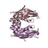
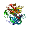
| Title | Crystal structure of a VirB8 Type IV secretion system machinery soluble domain from Bartonella tribocorum | ||||||
 Components Components | VirB8 protein | ||||||
 Keywords Keywords | PROTEIN TRANSPORT / Structural Genomics / NIAID / National Institute of Allergy and Infectious Diseases / Seattle Structural Genomics Center for Infectious Disease / SSGCID / transmembrane protein / type IV secretion system / T4SS | ||||||
| Function / homology |  Function and homology information Function and homology informationprotein secretion by the type IV secretion system / endomembrane system / membrane Similarity search - Function | ||||||
| Biological species |  Bartonella tribocorum (bacteria) Bartonella tribocorum (bacteria) | ||||||
| Method |  X-RAY DIFFRACTION / SYNCHROTRON / MOLECULAR REPLACEMENT / molecular replacement / Resolution: 2.85 Å X-RAY DIFFRACTION / SYNCHROTRON / MOLECULAR REPLACEMENT / molecular replacement / Resolution: 2.85 Å | ||||||
 Authors Authors | Seattle Structural Genomics Center for Infectious Disease (SSGCID) | ||||||
 Citation Citation | Journal: MBio / Year: 2015 Title: Structural Insight into How Bacteria Prevent Interference between Multiple Divergent Type IV Secretion Systems. Authors: Gillespie, J.J. / Phan, I.Q. / Scheib, H. / Subramanian, S. / Edwards, T.E. / Lehman, S.S. / Piitulainen, H. / Rahman, M.S. / Rennoll-Bankert, K.E. / Staker, B.L. / Taira, S. / Stacy, R. / ...Authors: Gillespie, J.J. / Phan, I.Q. / Scheib, H. / Subramanian, S. / Edwards, T.E. / Lehman, S.S. / Piitulainen, H. / Rahman, M.S. / Rennoll-Bankert, K.E. / Staker, B.L. / Taira, S. / Stacy, R. / Myler, P.J. / Azad, A.F. / Pulliainen, A.T. | ||||||
| History |
|
- Structure visualization
| Structure viewer | Molecule: MolmilJmol/JSmol |
|---|
- Downloads & links
-Download
| PDBx/mmCIF format | 4mei.cif.gz | 69.3 KB | Display | PDBx/mmCIF format |
|---|---|---|---|---|
| PDB format | pdb4mei.ent.gz | 51.1 KB | Display | PDB format |
| PDBx/mmJSON format | 4mei.json.gz | Tree view | PDBx/mmJSON format | |
| Others |  Other downloads Other downloads |
-Validation report
| Arichive directory | https://data.pdbj.org/pub/pdb/validation_reports/me/4meiftp://data.pdbj.org/pub/pdb/validation_reports/me/4mei | HTTPS FTP |
|---|
-Related structure data
| Related structure data |  4jf8C 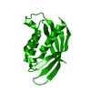 4kz1C  4lsoC  4nhfC 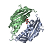 4o3vC C: citing same article ( |
|---|---|
| Similar structure data | |
| Other databases |


-Links
 PDBj
PDBj

- Assembly
| Deposited unit | 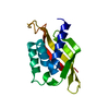
| ||||||||
|---|---|---|---|---|---|---|---|---|---|
| 1 | 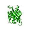
| ||||||||
| Unit cell |
|
-Components
| #1: Protein | Mass: 20837.367 Da / Num. of mol.: 1 / Fragment: UNP residues 51-222 Source method: isolated from a genetically manipulated source Source: (gene. exp.) Bartonella tribocorum (bacteria) / Strain: CIP 105476 / Gene: BT_1695, virB8 / Production host: |
|---|---|
| #2: Water | ChemComp-HOH /  Mass: 18.015 Da / Num. of mol.: 14 / Source method: isolated from a natural source / Formula: H2O Mass: 18.015 Da / Num. of mol.: 14 / Source method: isolated from a natural source / Formula: H2O |
-Experimental details
-Experiment
| Experiment | Method: X-RAY DIFFRACTION / Number of used crystals: 1 |
|---|
- Sample preparation
| Crystal | Density Matthews: 2.87 Å3/Da / Density % sol: 57.19 % |
|---|---|
| Crystal grow | Temperature: 289 K / Method: vapor diffusion, sitting drop / pH: 7 Details: BatrA.18388.b.B2.PS01799 at 3.84 mg/mL against JCSG+ F10 1.1 M Na Malonate, 0.1 M Hepes pH 7.0, 0.5% Jeffamine ED2001 with 20% ethylene glycol as cryo-protectant, crystal tracking ID ...Details: BatrA.18388.b.B2.PS01799 at 3.84 mg/mL against JCSG+ F10 1.1 M Na Malonate, 0.1 M Hepes pH 7.0, 0.5% Jeffamine ED2001 with 20% ethylene glycol as cryo-protectant, crystal tracking ID 244429f10, unique puck ID jcp2-9, VAPOR DIFFUSION, SITTING DROP, temperature 289K |
-Data collection
| Diffraction | Mean temperature: 100 K | |||||||||||||||||||||||||||||||||||||||||||||||||||||||||||||||||||||||||||||||||||||||||||||||||||||||||||||||||||||||||||||||||||||||||||||||||||
|---|---|---|---|---|---|---|---|---|---|---|---|---|---|---|---|---|---|---|---|---|---|---|---|---|---|---|---|---|---|---|---|---|---|---|---|---|---|---|---|---|---|---|---|---|---|---|---|---|---|---|---|---|---|---|---|---|---|---|---|---|---|---|---|---|---|---|---|---|---|---|---|---|---|---|---|---|---|---|---|---|---|---|---|---|---|---|---|---|---|---|---|---|---|---|---|---|---|---|---|---|---|---|---|---|---|---|---|---|---|---|---|---|---|---|---|---|---|---|---|---|---|---|---|---|---|---|---|---|---|---|---|---|---|---|---|---|---|---|---|---|---|---|---|---|---|---|---|---|
| Diffraction source | Source: SYNCHROTRON / Site: APS  / Beamline: 21-ID-G / Wavelength: 0.97856 Å / Beamline: 21-ID-G / Wavelength: 0.97856 Å | |||||||||||||||||||||||||||||||||||||||||||||||||||||||||||||||||||||||||||||||||||||||||||||||||||||||||||||||||||||||||||||||||||||||||||||||||||
| Detector | Type: RAYONIX MX-300 / Detector: CCD / Date: Jul 31, 2012 | |||||||||||||||||||||||||||||||||||||||||||||||||||||||||||||||||||||||||||||||||||||||||||||||||||||||||||||||||||||||||||||||||||||||||||||||||||
| Radiation | Protocol: SINGLE WAVELENGTH / Monochromatic (M) / Laue (L): M / Scattering type: x-ray | |||||||||||||||||||||||||||||||||||||||||||||||||||||||||||||||||||||||||||||||||||||||||||||||||||||||||||||||||||||||||||||||||||||||||||||||||||
| Radiation wavelength | Wavelength: 0.97856 Å / Relative weight: 1 | |||||||||||||||||||||||||||||||||||||||||||||||||||||||||||||||||||||||||||||||||||||||||||||||||||||||||||||||||||||||||||||||||||||||||||||||||||
| Reflection | Resolution: 2.85→50 Å / Num. all: 6185 / Num. obs: 6166 / % possible obs: 99.7 % / Observed criterion σ(I): -3 / Redundancy: 8.5 % / Biso Wilson estimate: 56.293 Å2 / Rmerge(I) obs: 0.077 / Net I/σ(I): 22.98 | |||||||||||||||||||||||||||||||||||||||||||||||||||||||||||||||||||||||||||||||||||||||||||||||||||||||||||||||||||||||||||||||||||||||||||||||||||
| Reflection shell | Diffraction-ID: 1
|
-Phasing
| Phasing | Method: molecular replacement | |||||||||
|---|---|---|---|---|---|---|---|---|---|---|
| Phasing MR | Model details: Phaser MODE: MR_AUTO
|
- Processing
| Software |
| |||||||||||||||||||||||||||||||||||||||||||||||||||||||||||||||||||||||||||
|---|---|---|---|---|---|---|---|---|---|---|---|---|---|---|---|---|---|---|---|---|---|---|---|---|---|---|---|---|---|---|---|---|---|---|---|---|---|---|---|---|---|---|---|---|---|---|---|---|---|---|---|---|---|---|---|---|---|---|---|---|---|---|---|---|---|---|---|---|---|---|---|---|---|---|---|---|
| Refinement | Method to determine structure: MOLECULAR REPLACEMENT / Resolution: 2.85→44.14 Å / Cor.coef. Fo:Fc: 0.936 / Cor.coef. Fo:Fc free: 0.904 / WRfactor Rfree: 0.2265 / WRfactor Rwork: 0.1812 / Occupancy max: 1 / Occupancy min: 0.5 / FOM work R set: 0.8377 / SU B: 22.597 / SU ML: 0.212 / SU R Cruickshank DPI: 0.5137 / SU Rfree: 0.3121 / Cross valid method: THROUGHOUT / σ(F): 0 / ESU R: 0.514 / ESU R Free: 0.312 / Stereochemistry target values: MAXIMUM LIKELIHOOD Details: U VALUES : WITH TLS ADDED HYDROGENS HAVE BEEN ADDED IN THE RIDING POSITIONS
| |||||||||||||||||||||||||||||||||||||||||||||||||||||||||||||||||||||||||||
| Solvent computation | Ion probe radii: 0.8 Å / Shrinkage radii: 0.8 Å / VDW probe radii: 1.2 Å / Solvent model: MASK | |||||||||||||||||||||||||||||||||||||||||||||||||||||||||||||||||||||||||||
| Displacement parameters | Biso max: 116.42 Å2 / Biso mean: 58.9129 Å2 / Biso min: 38.2 Å2
| |||||||||||||||||||||||||||||||||||||||||||||||||||||||||||||||||||||||||||
| Refinement step | Cycle: LAST / Resolution: 2.85→44.14 Å
| |||||||||||||||||||||||||||||||||||||||||||||||||||||||||||||||||||||||||||
| Refine LS restraints |
| |||||||||||||||||||||||||||||||||||||||||||||||||||||||||||||||||||||||||||
| LS refinement shell | Resolution: 2.85→2.924 Å / Total num. of bins used: 20
| |||||||||||||||||||||||||||||||||||||||||||||||||||||||||||||||||||||||||||
| Refinement TLS params. | Method: refined / Origin x: 8.2964 Å / Origin y: 46.394 Å / Origin z: 20.7892 Å
|
