 Movie
Movie Controller

[English] 日本語
 Yorodumi
- PDB-7s8s: Crystal Structure of HLA A*1101 in complex with RVLVNGTFLK, an 10... -
+ Open data

- Basic information
| Entry | Database: PDB / ID: 7s8s | ||||||
|---|---|---|---|---|---|---|---|
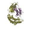
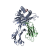
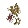
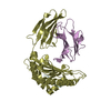
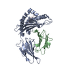
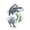
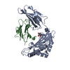
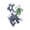
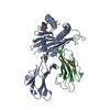
| Title | Crystal Structure of HLA A*1101 in complex with RVLVNGTFLK, an 10-mer epitope from Influenza B | ||||||
 Components Components |
| ||||||
 Keywords Keywords | IMMUNE SYSTEM / HLA A*1101 / influenza B / TCR / T cell | ||||||
| Function / homology |  Function and homology information Function and homology informationsymbiont-mediated suppression of host PKR/eIFalpha signaling / : / antigen processing and presentation of peptide antigen via MHC class I / protein serine/threonine kinase inhibitor activity / negative regulation of receptor binding / early endosome lumen / Nef mediated downregulation of MHC class I complex cell surface expression / DAP12 interactions / transferrin transport / cellular response to iron ion ...symbiont-mediated suppression of host PKR/eIFalpha signaling / : / antigen processing and presentation of peptide antigen via MHC class I / protein serine/threonine kinase inhibitor activity / negative regulation of receptor binding / early endosome lumen / Nef mediated downregulation of MHC class I complex cell surface expression / DAP12 interactions / transferrin transport / cellular response to iron ion / Endosomal/Vacuolar pathway / Antigen Presentation: Folding, assembly and peptide loading of class I MHC / peptide antigen assembly with MHC class II protein complex / lumenal side of endoplasmic reticulum membrane / cellular response to iron(III) ion / negative regulation of forebrain neuron differentiation / MHC class II protein complex / antigen processing and presentation of exogenous protein antigen via MHC class Ib, TAP-dependent / ER to Golgi transport vesicle membrane / peptide antigen assembly with MHC class I protein complex / regulation of iron ion transport / regulation of erythrocyte differentiation / HFE-transferrin receptor complex / response to molecule of bacterial origin / MHC class I peptide loading complex / T cell mediated cytotoxicity / positive regulation of T cell cytokine production / antigen processing and presentation of endogenous peptide antigen via MHC class I / antigen processing and presentation of exogenous peptide antigen via MHC class II / positive regulation of immune response / MHC class I protein complex / peptide antigen binding / positive regulation of T cell activation / positive regulation of receptor-mediated endocytosis / negative regulation of neurogenesis / cellular response to nicotine / positive regulation of T cell mediated cytotoxicity / multicellular organismal-level iron ion homeostasis / specific granule lumen / phagocytic vesicle membrane / recycling endosome membrane / Interferon gamma signaling / Immunoregulatory interactions between a Lymphoid and a non-Lymphoid cell / negative regulation of epithelial cell proliferation / MHC class II protein complex binding / Modulation by Mtb of host immune system / late endosome membrane / sensory perception of smell / positive regulation of cellular senescence / tertiary granule lumen / DAP12 signaling / T cell differentiation in thymus / negative regulation of neuron projection development / ER-Phagosome pathway / protein refolding / early endosome membrane / symbiont-mediated suppression of host ISG15-protein conjugation / protein homotetramerization / amyloid fibril formation / intracellular iron ion homeostasis / learning or memory / host cell cytoplasm / symbiont-mediated suppression of host type I interferon-mediated signaling pathway / endoplasmic reticulum lumen / Amyloid fiber formation / Golgi membrane / lysosomal membrane / external side of plasma membrane / focal adhesion / Neutrophil degranulation / host cell nucleus / SARS-CoV-2 activates/modulates innate and adaptive immune responses / structural molecule activity / endoplasmic reticulum / Golgi apparatus / protein homodimerization activity / extracellular space / RNA binding / extracellular exosome / extracellular region / identical protein binding / membrane / plasma membrane / cytosol Similarity search - Function | ||||||
| Biological species |  Homo sapiens (human) Homo sapiens (human) Influenza B virus Influenza B virus | ||||||
| Method |  X-RAY DIFFRACTION / SYNCHROTRON / MOLECULAR REPLACEMENT / Resolution: 1.87 Å X-RAY DIFFRACTION / SYNCHROTRON / MOLECULAR REPLACEMENT / Resolution: 1.87 Å | ||||||
 Authors Authors | Nguyen, A.T. / Szeto, C. / Gras, S. | ||||||
| Funding support |  Australia, 1items Australia, 1items
| ||||||
 Citation Citation | Journal: Plos Pathog. / Year: 2022 Title: HLA-A*11:01-restricted CD8+ T cell immunity against influenza A and influenza B viruses in Indigenous and non-Indigenous people. Authors: Habel, J.R. / Nguyen, A.T. / Rowntree, L.C. / Szeto, C. / Mifsud, N.A. / Clemens, E.B. / Loh, L. / Chen, W. / Rockman, S. / Nelson, J. / Davies, J. / Miller, A. / Tong, S.Y.C. / Rossjohn, J. ...Authors: Habel, J.R. / Nguyen, A.T. / Rowntree, L.C. / Szeto, C. / Mifsud, N.A. / Clemens, E.B. / Loh, L. / Chen, W. / Rockman, S. / Nelson, J. / Davies, J. / Miller, A. / Tong, S.Y.C. / Rossjohn, J. / Gras, S. / Purcell, A.W. / Hensen, L. / Kedzierska, K. / Illing, P.T. | ||||||
| History |
|
- Structure visualization
| Structure viewer | Molecule: MolmilJmol/JSmol |
|---|
- Downloads & links
-Download
| PDBx/mmCIF format | 7s8s.cif.gz | 99.6 KB | Display | PDBx/mmCIF format |
|---|---|---|---|---|
| PDB format | pdb7s8s.ent.gz | 73.6 KB | Display | PDB format |
| PDBx/mmJSON format | 7s8s.json.gz | Tree view | PDBx/mmJSON format | |
| Others |  Other downloads Other downloads |
-Validation report
| Summary document | 7s8s_validation.pdf.gz | 448.3 KB | Display | wwPDB validaton report |
|---|---|---|---|---|
| Full document | 7s8s_full_validation.pdf.gz | 450 KB | Display | |
| Data in XML | 7s8s_validation.xml.gz | 17.8 KB | Display | |
| Data in CIF | 7s8s_validation.cif.gz | 25.6 KB | Display | |
| Arichive directory | https://data.pdbj.org/pub/pdb/validation_reports/s8/7s8sftp://data.pdbj.org/pub/pdb/validation_reports/s8/7s8s | HTTPS FTP |
-Related structure data
| Related structure data | 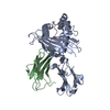 7s8qC  7s8rC  4mj5S S: Starting model for refinement C: citing same article ( |
|---|---|
| Similar structure data |


-Links
 PDBj
PDBj

- Assembly
| Deposited unit | 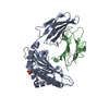
| ||||||||
|---|---|---|---|---|---|---|---|---|---|
| 1 |
| ||||||||
| Unit cell |
|
-Components
| #1: Protein | Mass: 32142.363 Da / Num. of mol.: 1 Source method: isolated from a genetically manipulated source Source: (gene. exp.) Homo sapiens (human) / Gene: HLA-A / Plasmid: pET / Production host:  |
|---|---|
| #2: Protein | Mass: 11879.356 Da / Num. of mol.: 1 / Fragment: UNP residues 21-119 Source method: isolated from a genetically manipulated source Source: (gene. exp.) Homo sapiens (human) / Gene: B2M, CDABP0092, HDCMA22P / Plasmid: pET / Production host: |
| #3: Protein/peptide | Mass: 1148.398 Da / Num. of mol.: 1 / Fragment: UNP residues 186-195 / Source method: obtained synthetically / Details: peptide from Influenza B / Source: (synth.) Influenza B virus / References: UniProt: P12601 |
| #4: Chemical | ChemComp-SO4 /   Mass: 96.063 Da / Num. of mol.: 1 / Source method: obtained synthetically / Formula: SO4 Mass: 96.063 Da / Num. of mol.: 1 / Source method: obtained synthetically / Formula: SO4 |
| #5: Water | ChemComp-HOH /  Mass: 18.015 Da / Num. of mol.: 230 / Source method: isolated from a natural source / Formula: H2O Mass: 18.015 Da / Num. of mol.: 230 / Source method: isolated from a natural source / Formula: H2O |
| Has ligand of interest | N |
| Has protein modification | Y |
-Experimental details
-Experiment
| Experiment | Method: X-RAY DIFFRACTION / Number of used crystals: 1 |
|---|
- Sample preparation
| Crystal | Density Matthews: 2.43 Å3/Da / Density % sol: 49.41 % / Mosaicity: 0.18 ° |
|---|---|
| Crystal grow | Temperature: 277 K / Method: vapor diffusion, sitting drop / pH: 6.5 Details: 26% PEG 3350, 0.1M BisTris pH 6.3, 0.2M Lithium Sulfate |
-Data collection
| Diffraction | Mean temperature: 100 K / Serial crystal experiment: N |
|---|---|
| Diffraction source | Source: SYNCHROTRON / Site: Australian Synchrotron / Beamline: MX2 / Wavelength: 0.98 Å |
| Detector | Type: DECTRIS PILATUS 12M / Detector: PIXEL / Date: Oct 17, 2020 |
| Radiation | Protocol: SINGLE WAVELENGTH / Monochromatic (M) / Laue (L): M / Scattering type: x-ray |
| Radiation wavelength | Wavelength: 0.98 Å / Relative weight: 1 |
| Reflection | Resolution: 1.82→46.82 Å / Num. obs: 38748 / % possible obs: 99.9 % / Redundancy: 6.8 % / Biso Wilson estimate: 26.21 Å2 / CC1/2: 0.997 / Rmerge(I) obs: 0.125 / Rpim(I) all: 0.052 / Rrim(I) all: 0.135 / Net I/σ(I): 9.9 / Num. measured all: 265223 / Scaling rejects: 113 |
| Reflection shell | Resolution: 1.82→1.86 Å / Redundancy: 7 % / Rmerge(I) obs: 1.183 / Num. unique obs: 2267 / CC1/2: 0.682 / Rpim(I) all: 0.481 / % possible all: 100 |
- Processing
| Software |
| ||||||||||||||||||||||||||||||||||||||||||||||||||||||||||||||||||||||||||||||||||||||||||||||||||||||||||||
|---|---|---|---|---|---|---|---|---|---|---|---|---|---|---|---|---|---|---|---|---|---|---|---|---|---|---|---|---|---|---|---|---|---|---|---|---|---|---|---|---|---|---|---|---|---|---|---|---|---|---|---|---|---|---|---|---|---|---|---|---|---|---|---|---|---|---|---|---|---|---|---|---|---|---|---|---|---|---|---|---|---|---|---|---|---|---|---|---|---|---|---|---|---|---|---|---|---|---|---|---|---|---|---|---|---|---|---|---|---|
| Refinement | Method to determine structure: MOLECULAR REPLACEMENT Starting model: 4MJ5 Resolution: 1.87→25.39 Å / Cor.coef. Fo:Fc: 0.927 / Cor.coef. Fo:Fc free: 0.912 / SU R Cruickshank DPI: 0.142 / Cross valid method: THROUGHOUT / σ(F): 0 / SU R Blow DPI: 0.143 / SU Rfree Blow DPI: 0.13 / SU Rfree Cruickshank DPI: 0.13
| ||||||||||||||||||||||||||||||||||||||||||||||||||||||||||||||||||||||||||||||||||||||||||||||||||||||||||||
| Displacement parameters | Biso max: 98.71 Å2 / Biso mean: 29.98 Å2 / Biso min: 12.28 Å2
| ||||||||||||||||||||||||||||||||||||||||||||||||||||||||||||||||||||||||||||||||||||||||||||||||||||||||||||
| Refine analyze | Luzzati coordinate error obs: 0.25 Å | ||||||||||||||||||||||||||||||||||||||||||||||||||||||||||||||||||||||||||||||||||||||||||||||||||||||||||||
| Refinement step | Cycle: final / Resolution: 1.87→25.39 Å
| ||||||||||||||||||||||||||||||||||||||||||||||||||||||||||||||||||||||||||||||||||||||||||||||||||||||||||||
| Refine LS restraints |
| ||||||||||||||||||||||||||||||||||||||||||||||||||||||||||||||||||||||||||||||||||||||||||||||||||||||||||||
| LS refinement shell | Resolution: 1.87→1.88 Å / Rfactor Rfree error: 0 / Total num. of bins used: 50
|
