 Movie
Movie Controller

[English] 日本語
 Yorodumi
- PDB-7s4y: Serial Macromolecular Crystallography at ALBA Synchrotron Light S... -
+ Open data

- Basic information
| Entry | Database: PDB / ID: 7s4y | |||||||||
|---|---|---|---|---|---|---|---|---|---|---|
| Title | Serial Macromolecular Crystallography at ALBA Synchrotron Light Source - Insulin | |||||||||
 Components Components |
| |||||||||
 Keywords Keywords | HORMONE / Serial Crystallography | |||||||||
| Function / homology |  Function and homology information Function and homology informationnegative regulation of glycogen catabolic process / : / negative regulation of fatty acid metabolic process / Signaling by Insulin receptor / negative regulation of feeding behavior / IRS activation / Insulin processing / regulation of protein secretion / positive regulation of peptide hormone secretion / negative regulation of acute inflammatory response ...negative regulation of glycogen catabolic process / : / negative regulation of fatty acid metabolic process / Signaling by Insulin receptor / negative regulation of feeding behavior / IRS activation / Insulin processing / regulation of protein secretion / positive regulation of peptide hormone secretion / negative regulation of acute inflammatory response / Regulation of gene expression in beta cells / positive regulation of respiratory burst / alpha-beta T cell activation / Synthesis, secretion, and deacylation of Ghrelin / negative regulation of protein secretion / positive regulation of dendritic spine maintenance / negative regulation of gluconeogenesis / fatty acid homeostasis / positive regulation of glycogen biosynthetic process / positive regulation of insulin receptor signaling pathway / Signal attenuation / FOXO-mediated transcription of oxidative stress, metabolic and neuronal genes / negative regulation of respiratory burst involved in inflammatory response / negative regulation of lipid catabolic process / positive regulation of lipid biosynthetic process / negative regulation of oxidative stress-induced intrinsic apoptotic signaling pathway / nitric oxide-cGMP-mediated signaling / regulation of protein localization to plasma membrane / positive regulation of nitric-oxide synthase activity / transport vesicle / Insulin receptor recycling / COPI-mediated anterograde transport / positive regulation of brown fat cell differentiation / negative regulation of reactive oxygen species biosynthetic process / insulin-like growth factor receptor binding / NPAS4 regulates expression of target genes / neuron projection maintenance / positive regulation of mitotic nuclear division / endoplasmic reticulum-Golgi intermediate compartment membrane / Insulin receptor signalling cascade / positive regulation of glycolytic process / endosome lumen / positive regulation of cytokine production / acute-phase response / positive regulation of D-glucose import across plasma membrane / insulin receptor binding / positive regulation of long-term synaptic potentiation / positive regulation of protein secretion / positive regulation of cell differentiation / wound healing / Regulation of insulin secretion / hormone activity / positive regulation of neuron projection development / negative regulation of protein catabolic process / positive regulation of protein localization to nucleus / regulation of synaptic plasticity / Golgi lumen / cognition / vasodilation / glucose metabolic process / insulin receptor signaling pathway / cell-cell signaling / regulation of protein localization / glucose homeostasis / PI5P, PP2A and IER3 Regulate PI3K/AKT Signaling / positive regulation of cell growth / protease binding / secretory granule lumen / positive regulation of MAPK cascade / positive regulation of canonical NF-kappaB signal transduction / positive regulation of phosphatidylinositol 3-kinase/protein kinase B signal transduction / positive regulation of cell migration / endoplasmic reticulum lumen / G protein-coupled receptor signaling pathway / Amyloid fiber formation / receptor ligand activity / Golgi membrane / negative regulation of gene expression / positive regulation of cell population proliferation / positive regulation of gene expression / regulation of DNA-templated transcription / : / extracellular region / identical protein binding Similarity search - Function | |||||||||
| Biological species |  Homo sapiens (human) Homo sapiens (human) | |||||||||
| Method |  X-RAY DIFFRACTION / SYNCHROTRON / MOLECULAR REPLACEMENT / Resolution: 1.71 Å X-RAY DIFFRACTION / SYNCHROTRON / MOLECULAR REPLACEMENT / Resolution: 1.71 Å | |||||||||
 Authors Authors | Martin-Garcia, J.M. / Botha, S. / Hu, H. / Jernigan, R. / Castellvi, A. / Lisova, S. / Gil, F. / Calisto, B. / Crespo, I. / Roy-Chowdbury, S. ...Martin-Garcia, J.M. / Botha, S. / Hu, H. / Jernigan, R. / Castellvi, A. / Lisova, S. / Gil, F. / Calisto, B. / Crespo, I. / Roy-Chowdbury, S. / Grieco, A. / Ketawala, G. / Weierstall, U. / Spence, J. / Fromme, P. / Zatsepin, N. / Boer, R. / Carpena, X. | |||||||||
| Funding support | 2items
| |||||||||
 Citation Citation | Journal: J.Synchrotron Radiat. / Year: 2022 Title: Serial macromolecular crystallography at ALBA Synchrotron Light Source. Authors: Martin-Garcia, J.M. / Botha, S. / Hu, H. / Jernigan, R. / Castellvi, A. / Lisova, S. / Gil, F. / Calisto, B. / Crespo, I. / Roy-Chowdhury, S. / Grieco, A. / Ketawala, G. / Weierstall, U. / ...Authors: Martin-Garcia, J.M. / Botha, S. / Hu, H. / Jernigan, R. / Castellvi, A. / Lisova, S. / Gil, F. / Calisto, B. / Crespo, I. / Roy-Chowdhury, S. / Grieco, A. / Ketawala, G. / Weierstall, U. / Spence, J. / Fromme, P. / Zatsepin, N. / Boer, D.R. / Carpena, X. | |||||||||
| History |
|
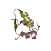
- Structure visualization
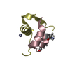
| Structure viewer | Molecule: MolmilJmol/JSmol |
|---|
- Downloads & links
-Download
| PDBx/mmCIF format | 7s4y.cif.gz | 34.6 KB | Display | PDBx/mmCIF format |
|---|---|---|---|---|
| PDB format | pdb7s4y.ent.gz | 22.1 KB | Display | PDB format |
| PDBx/mmJSON format | 7s4y.json.gz | Tree view | PDBx/mmJSON format | |
| Others |  Other downloads Other downloads |
-Validation report
| Arichive directory | https://data.pdbj.org/pub/pdb/validation_reports/s4/7s4yftp://data.pdbj.org/pub/pdb/validation_reports/s4/7s4y | HTTPS FTP |
|---|
-Related structure data
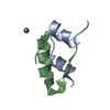
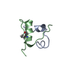
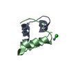
| Related structure data |  7s4rC  7s4wC  7s4zC 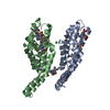 7s50C  4ey9S C: citing same article ( S: Starting model for refinement |
|---|---|
| Similar structure data |

-Links
 PDBj
PDBj


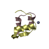
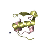
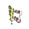
- Assembly
| Deposited unit | 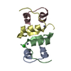
| ||||||||
|---|---|---|---|---|---|---|---|---|---|
| 1 | 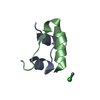
| ||||||||
| 2 | 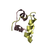
| ||||||||
| Unit cell |
| ||||||||
| Components on special symmetry positions |
|
-Components
| #1: Protein/peptide | Mass: 2383.698 Da / Num. of mol.: 2 Source method: isolated from a genetically manipulated source Source: (gene. exp.) Homo sapiens (human) / Gene: INS / Production host: Homo sapiens (human) / References: UniProt: P01308#2: Protein/peptide | Mass: 3433.953 Da / Num. of mol.: 2 Source method: isolated from a genetically manipulated source Source: (gene. exp.) Homo sapiens (human) / Gene: INS / Production host: Homo sapiens (human) / References: UniProt: P01308#3: Chemical |   Mass: 65.409 Da / Num. of mol.: 2 / Source method: obtained synthetically / Formula: Zn Mass: 65.409 Da / Num. of mol.: 2 / Source method: obtained synthetically / Formula: Zn#4: Chemical | ChemComp-CL / |   Mass: 35.453 Da / Num. of mol.: 1 / Source method: obtained synthetically / Formula: Cl Mass: 35.453 Da / Num. of mol.: 1 / Source method: obtained synthetically / Formula: Cl#5: Water | ChemComp-HOH / |  Mass: 18.015 Da / Num. of mol.: 15 / Source method: isolated from a natural source / Formula: H2O Mass: 18.015 Da / Num. of mol.: 15 / Source method: isolated from a natural source / Formula: H2OHas ligand of interest | N | Has protein modification | Y | |
|---|
-Experimental details
-Experiment
| Experiment | Method: X-RAY DIFFRACTION / Number of used crystals: 1 |
|---|
- Sample preparation
| Crystal | Density Matthews: 1.85 Å3/Da / Density % sol: 33.52 % |
|---|---|
| Crystal grow | Temperature: 298 K / Method: batch mode Details: insulin was dissolved in 5 mM zinc chloride, 25 mM HCl at a concentration of 5-10 mg/ml. Cuboid-shaped microcrystals were obtained in 35.2 mM sodium citrate pH 7, and 5% (v/v) acetone as precipitant |
-Data collection
| Diffraction | Mean temperature: 298 K / Serial crystal experiment: Y |
|---|---|
| Diffraction source | Source: SYNCHROTRON / Site: ALBA  / Beamline: XALOC / Wavelength: 1.27819 Å / Beamline: XALOC / Wavelength: 1.27819 Å |
| Detector | Type: DECTRIS PILATUS 6M / Detector: PIXEL / Date: Jun 8, 2019 |
| Radiation | Protocol: SINGLE WAVELENGTH / Monochromatic (M) / Laue (L): M / Scattering type: x-ray |
| Radiation wavelength | Wavelength: 1.27819 Å / Relative weight: 1 |
| Reflection | Resolution: 1.7→30.3 Å / Num. obs: 9140 / % possible obs: 100 % / Redundancy: 1292 % / Biso Wilson estimate: 29.1 Å2 / CC1/2: 0.9789 / CC star: 0.9947 / Net I/σ(I): 7.9 |
| Reflection shell | Resolution: 1.7→1.76 Å / Redundancy: 881 % / Mean I/σ(I) obs: 1.5 / Num. unique obs: 928 / CC1/2: 0.36711 / CC star: 0.7328 / % possible all: 100 |
| Serial crystallography sample delivery | Description: LCP Injector / Method: injection |
| Serial crystallography sample delivery injection | Injector diameter: 5.0E-5 µm |
| Serial crystallography data reduction | Frames total: 433986 / Lattices indexed: 51144 |
- Processing
| Software |
| |||||||||||||||||||||||||||||||||||||||||||||||||||||||||||||||||||||||||||||||||||||||||||||||||||||||||
|---|---|---|---|---|---|---|---|---|---|---|---|---|---|---|---|---|---|---|---|---|---|---|---|---|---|---|---|---|---|---|---|---|---|---|---|---|---|---|---|---|---|---|---|---|---|---|---|---|---|---|---|---|---|---|---|---|---|---|---|---|---|---|---|---|---|---|---|---|---|---|---|---|---|---|---|---|---|---|---|---|---|---|---|---|---|---|---|---|---|---|---|---|---|---|---|---|---|---|---|---|---|---|---|---|---|---|
| Refinement | Method to determine structure: MOLECULAR REPLACEMENT Starting model: 4EY9 Resolution: 1.71→24.35 Å / Cor.coef. Fo:Fc: 0.935 / Cor.coef. Fo:Fc free: 0.91 / SU B: 2.882 / SU ML: 0.102 / Cross valid method: THROUGHOUT / ESU R: 0.036 / ESU R Free: 0.03 / Stereochemistry target values: MAXIMUM LIKELIHOOD Details: HYDROGENS HAVE BEEN ADDED IN THE RIDING POSITIONS U VALUES : REFINED INDIVIDUALLY
| |||||||||||||||||||||||||||||||||||||||||||||||||||||||||||||||||||||||||||||||||||||||||||||||||||||||||
| Solvent computation | Ion probe radii: 0.9 Å / Shrinkage radii: 0.9 Å / VDW probe radii: 1 Å / Solvent model: MASK | |||||||||||||||||||||||||||||||||||||||||||||||||||||||||||||||||||||||||||||||||||||||||||||||||||||||||
| Displacement parameters | Biso mean: 33.509 Å2
| |||||||||||||||||||||||||||||||||||||||||||||||||||||||||||||||||||||||||||||||||||||||||||||||||||||||||
| Refinement step | Cycle: LAST / Resolution: 1.71→24.35 Å
| |||||||||||||||||||||||||||||||||||||||||||||||||||||||||||||||||||||||||||||||||||||||||||||||||||||||||
| Refine LS restraints |
| |||||||||||||||||||||||||||||||||||||||||||||||||||||||||||||||||||||||||||||||||||||||||||||||||||||||||
| LS refinement shell | Resolution: 1.71→1.749 Å
|
