 Movie
Movie Controller

[English] 日本語
 Yorodumi
- PDB-7rxn: STRUCTURE OF RUBREDOXIN FROM DESULFOVIBRIO VULGARIS AT 1.5 A RESO... -
+ Open data

- Basic information
| Entry | Database: PDB / ID: 7rxn | |||||||||
|---|---|---|---|---|---|---|---|---|---|---|
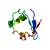
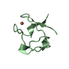
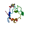
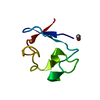
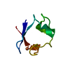
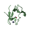
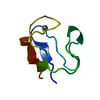
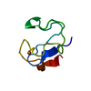
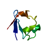
| Title | STRUCTURE OF RUBREDOXIN FROM DESULFOVIBRIO VULGARIS AT 1.5 A RESOLUTION | |||||||||
 Components Components | RUBREDOXIN | |||||||||
 Keywords Keywords | ELECTRON TRANSFER(IRON-SULFUR PROTEIN) | |||||||||
| Function / homology |  Function and homology information Function and homology informationalkane catabolic process / electron transfer activity / iron ion binding / cytoplasm Similarity search - Function | |||||||||
| Biological species |  Desulfovibrio vulgaris (bacteria) Desulfovibrio vulgaris (bacteria) | |||||||||
| Method |  X-RAY DIFFRACTION / Resolution: 1.5 Å X-RAY DIFFRACTION / Resolution: 1.5 Å | |||||||||
 Authors Authors | Adman, E.T. / Sieker, L.C. / Jensen, L.H. | |||||||||
 Citation Citation | Journal: J.Mol.Biol. / Year: 1991 Title: Structure of rubredoxin from Desulfovibrio vulgaris at 1.5 A resolution. Authors: Adman, E.T. / Sieker, L.C. / Jensen, L.H. #1: Journal: Am.Cryst.Assoc.,Abstr.Papers (Winter Meeting) / Year: 1979Title: Progress on Refinement of Rubredoxin (D.Vulgaris) at 1.5 Angstroms Authors: Adman, E.T. / Jensen, L.H. #2: Journal: J.Mol.Biol. / Year: 1977Title: A Structural Model of Rubredoxin from Desulfovibrio Vulgaris at 2 Angstroms Resolution Authors: Adman, E.T. / Sieker, L.C. / Jensen, L.H. / Bruschi, M. / Legall, J. | |||||||||
| History |
|
- Structure visualization
| Structure viewer | Molecule: MolmilJmol/JSmol |
|---|
- Downloads & links
-Download
| PDBx/mmCIF format | 7rxn.cif.gz | 24 KB | Display | PDBx/mmCIF format |
|---|---|---|---|---|
| PDB format | pdb7rxn.ent.gz | 14.6 KB | Display | PDB format |
| PDBx/mmJSON format | 7rxn.json.gz | Tree view | PDBx/mmJSON format | |
| Others |  Other downloads Other downloads |
-Validation report
| Arichive directory | https://data.pdbj.org/pub/pdb/validation_reports/rx/7rxnftp://data.pdbj.org/pub/pdb/validation_reports/rx/7rxn | HTTPS FTP |
|---|
-Related structure data
| Similar structure data |
|---|

-Links
 PDBj
PDBj
- Assembly
| Deposited unit | 
| ||||||||
|---|---|---|---|---|---|---|---|---|---|
| 1 |
| ||||||||
| Unit cell |
|
-Components
| #1: Protein | Mass: 5578.174 Da / Num. of mol.: 1 Source method: isolated from a genetically manipulated source Source: (gene. exp.) Desulfovibrio vulgaris (bacteria) / References: UniProt: P00269 |
|---|---|
| #2: Chemical | ChemComp-FE /   Mass: 55.845 Da / Num. of mol.: 1 / Source method: obtained synthetically / Formula: Fe Mass: 55.845 Da / Num. of mol.: 1 / Source method: obtained synthetically / Formula: Fe |
| #3: Chemical | ChemComp-SO4 /   Mass: 96.063 Da / Num. of mol.: 1 / Source method: obtained synthetically / Formula: SO4 Mass: 96.063 Da / Num. of mol.: 1 / Source method: obtained synthetically / Formula: SO4 |
| #4: Water | ChemComp-HOH /  Mass: 18.015 Da / Num. of mol.: 180 / Source method: isolated from a natural source / Formula: H2O Mass: 18.015 Da / Num. of mol.: 180 / Source method: isolated from a natural source / Formula: H2O |
-Experimental details
-Experiment
| Experiment | Method: X-RAY DIFFRACTION |
|---|
- Sample preparation
| Crystal | Density Matthews: 1.73 Å3/Da / Density % sol: 28.91 % | ||||||||||||||||||||||||
|---|---|---|---|---|---|---|---|---|---|---|---|---|---|---|---|---|---|---|---|---|---|---|---|---|---|
| Crystal grow | *PLUS pH: 4 / Method: unknown | ||||||||||||||||||||||||
| Components of the solutions | *PLUS
|
-Data collection
| Reflection | *PLUS Highest resolution: 1.5 Å / Lowest resolution: 5 Å / Observed criterion σ(F): 2 |
|---|
- Processing
| Software | Name: PROLSQ / Classification: refinement | ||||||||||||||||||||||||||||||||||||||||||||||||||||||||||||||||||||||||||||||||||||
|---|---|---|---|---|---|---|---|---|---|---|---|---|---|---|---|---|---|---|---|---|---|---|---|---|---|---|---|---|---|---|---|---|---|---|---|---|---|---|---|---|---|---|---|---|---|---|---|---|---|---|---|---|---|---|---|---|---|---|---|---|---|---|---|---|---|---|---|---|---|---|---|---|---|---|---|---|---|---|---|---|---|---|---|---|---|
| Refinement | Rfactor obs: 0.098 / Highest resolution: 1.5 Å | ||||||||||||||||||||||||||||||||||||||||||||||||||||||||||||||||||||||||||||||||||||
| Refinement step | Cycle: LAST / Highest resolution: 1.5 Å
| ||||||||||||||||||||||||||||||||||||||||||||||||||||||||||||||||||||||||||||||||||||
| Refine LS restraints |
| ||||||||||||||||||||||||||||||||||||||||||||||||||||||||||||||||||||||||||||||||||||
| Software | *PLUS Name: 'PROLSQ, PROTIN' / Classification: refinement | ||||||||||||||||||||||||||||||||||||||||||||||||||||||||||||||||||||||||||||||||||||
| Refinement | *PLUS Lowest resolution: 9999 Å / Num. reflection obs: 5707 / Rfactor obs: 0.098 | ||||||||||||||||||||||||||||||||||||||||||||||||||||||||||||||||||||||||||||||||||||
| Solvent computation | *PLUS | ||||||||||||||||||||||||||||||||||||||||||||||||||||||||||||||||||||||||||||||||||||
| Displacement parameters | *PLUS |
