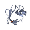
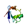
 Movie
Movie Controller

[English] 日本語
 Yorodumi
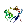
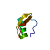
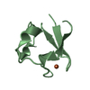
- PDB-1iu5: X-ray Crystal Structure of the rubredoxin mutant from Pyrococcus ... -
+ Open data

- Basic information
| Entry | Database: PDB / ID: 1iu5 | ||||||
|---|---|---|---|---|---|---|---|
| Title | X-ray Crystal Structure of the rubredoxin mutant from Pyrococcus Furiosus | ||||||
 Components Components | rubredoxin | ||||||
 Keywords Keywords | ELECTRON TRANSPORT / rubredoxin / mutant / thermostability | ||||||
| Function / homology |  Function and homology information Function and homology informationalkane catabolic process / electron transfer activity / iron ion binding Similarity search - Function | ||||||
| Biological species |   Pyrococcus furiosus (archaea) Pyrococcus furiosus (archaea) | ||||||
| Method |  X-RAY DIFFRACTION / MOLECULAR REPLACEMENT / Resolution: 1.5 Å X-RAY DIFFRACTION / MOLECULAR REPLACEMENT / Resolution: 1.5 Å | ||||||
 Authors Authors | Chatake, T. / Kurihara, K. / Tanaka, I. / Tsyba, I. / Bau, R. / Jenney, F.E. / Adams, M.W.W. / Niimura, N. | ||||||
 Citation Citation | Journal: Acta Crystallogr.,Sect.D / Year: 2004 Title: A neutron crystallographic analysis of a rubredoxin mutant at 1.6 A resolution. Authors: Chatake, T. / Kurihara, K. / Tanaka, I. / Tsyba, I. / Bau, R. / Jenney, F.E. / Adams, M.W. / Niimura, N. | ||||||
| History |
|
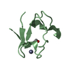
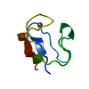
- Structure visualization
| Structure viewer | Molecule: MolmilJmol/JSmol |
|---|
- Downloads & links
-Download
| PDBx/mmCIF format | 1iu5.cif.gz | 22.6 KB | Display | PDBx/mmCIF format |
|---|---|---|---|---|
| PDB format | pdb1iu5.ent.gz | 13.4 KB | Display | PDB format |
| PDBx/mmJSON format | 1iu5.json.gz | Tree view | PDBx/mmJSON format | |
| Others |  Other downloads Other downloads |
-Validation report
| Arichive directory | https://data.pdbj.org/pub/pdb/validation_reports/iu/1iu5ftp://data.pdbj.org/pub/pdb/validation_reports/iu/1iu5 | HTTPS FTP |
|---|
-Related structure data
| Related structure data |  1iu6C  1brfS C: citing same article ( S: Starting model for refinement |
|---|---|
| Similar structure data |


-Links
 PDBj
PDBj


- Assembly
| Deposited unit | 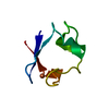
| ||||||||
|---|---|---|---|---|---|---|---|---|---|
| 1 |
| ||||||||
| Unit cell |
|
-Components
| #1: Protein | Mass: 5863.470 Da / Num. of mol.: 1 / Mutation: W3Y, I23V, L32I Source method: isolated from a genetically manipulated source Source: (gene. exp.) Pyrococcus furiosus (archaea) / Production host:  |
|---|---|
| #2: Chemical | ChemComp-FE /   Mass: 55.845 Da / Num. of mol.: 1 / Source method: obtained synthetically / Formula: Fe Mass: 55.845 Da / Num. of mol.: 1 / Source method: obtained synthetically / Formula: Fe |
| #3: Chemical | ChemComp-DOD /   Mass: 18.015 Da / Num. of mol.: 53 / Source method: isolated from a natural source / Formula: D2O Mass: 18.015 Da / Num. of mol.: 53 / Source method: isolated from a natural source / Formula: D2O |
-Experimental details
-Experiment
| Experiment | Method: X-RAY DIFFRACTION / Number of used crystals: 1 |
|---|
- Sample preparation
| Crystal | Density Matthews: 2.26 Å3/Da / Density % sol: 45.65 % | ||||||||||||||||||||||||||||||
|---|---|---|---|---|---|---|---|---|---|---|---|---|---|---|---|---|---|---|---|---|---|---|---|---|---|---|---|---|---|---|---|
| Crystal grow | Method: vapor diffusion / Details: Na/K phosphate, VAPOR DIFFUSION | ||||||||||||||||||||||||||||||
| Crystal grow | *PLUS pH: 8 / Method: vapor diffusion, hanging dropDetails: used macroseeding, Bau, R., (1998) J.BIOL.INORG.CHEM., 3, 484. | ||||||||||||||||||||||||||||||
| Components of the solutions | *PLUS
|
-Data collection
| Diffraction | Mean temperature: 293 K |
|---|---|
| Diffraction source | Source: ROTATING ANODE / Type: MACSCIENCE / Wavelength: 1.5418 Å |
| Detector | Type: MAC Science DIP-2000 / Detector: IMAGE PLATE / Date: May 29, 2001 |
| Radiation | Monochromator: graphite / Protocol: SINGLE WAVELENGTH / Monochromatic (M) / Laue (L): M / Scattering type: x-ray |
| Radiation wavelength | Wavelength: 1.5418 Å / Relative weight: 1 |
| Reflection | Resolution: 1.5→50 Å / Num. all: 8876 / Num. obs: 8876 / % possible obs: 98.6 % / Redundancy: 4.4 % / Biso Wilson estimate: 13.9 Å2 / Rmerge(I) obs: 0.077 / Net I/σ(I): 25.2 |
| Reflection shell | Resolution: 1.5→1.55 Å / Redundancy: 4.3 % / Rmerge(I) obs: 0.19 / % possible all: 97.7 |
| Reflection | *PLUS Num. measured all: 38773 |
| Reflection shell | *PLUS % possible obs: 97.7 % / Rmerge(I) obs: 0.19 |
- Processing
| Software |
| ||||||||||||||||
|---|---|---|---|---|---|---|---|---|---|---|---|---|---|---|---|---|---|
| Refinement | Method to determine structure: MOLECULAR REPLACEMENT Starting model: PDB ENTRY 1BRF Resolution: 1.5→10 Å / Isotropic thermal model: isotropic / σ(F): 3
| ||||||||||||||||
| Displacement parameters | Biso mean: 15.9 Å2 | ||||||||||||||||
| Refine analyze |
| ||||||||||||||||
| Refinement step | Cycle: LAST / Resolution: 1.5→10 Å
| ||||||||||||||||
| Refine LS restraints |
| ||||||||||||||||
| LS refinement shell | Resolution: 1.5→1.59 Å
| ||||||||||||||||
| Refinement | *PLUS Lowest resolution: 10 Å / Rfactor Rfree: 0.204 / Rfactor Rwork: 0.189 | ||||||||||||||||
| Solvent computation | *PLUS | ||||||||||||||||
| Displacement parameters | *PLUS |
