 Movie
Movie Controller

[English] 日本語
 Yorodumi
- PDB-7rw2: Cryo-EM structure of NTD-directed neutralizing antibody 5-7 in co... -
+ Open data

- Basic information
| Entry | Database: PDB / ID: 7rw2 | |||||||||
|---|---|---|---|---|---|---|---|---|---|---|
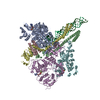
| Title | Cryo-EM structure of NTD-directed neutralizing antibody 5-7 in complex with prefusion SARS-CoV-2 spike glycoprotein | |||||||||
 Components Components |
| |||||||||
 Keywords Keywords | VIRAL PROTEIN/IMMUNE SYSTEM / Neutralizing antibody / Fusion protein / Spike glycoprotein / COVID-19 / N-terminal domain / NTD / 5-7 / VIRAL PROTEIN-IMMUNE SYSTEM complex | |||||||||
| Function / homology |  Function and homology information Function and homology informationsymbiont-mediated disruption of host tissue / Maturation of spike protein / Translation of Structural Proteins / Virion Assembly and Release / host cell surface / host extracellular region / symbiont-mediated-mediated suppression of host tetherin activity / Induction of Cell-Cell Fusion / structural constituent of virion / positive regulation of viral entry into host cell ...symbiont-mediated disruption of host tissue / Maturation of spike protein / Translation of Structural Proteins / Virion Assembly and Release / host cell surface / host extracellular region / symbiont-mediated-mediated suppression of host tetherin activity / Induction of Cell-Cell Fusion / structural constituent of virion / positive regulation of viral entry into host cell / membrane fusion / host cell endoplasmic reticulum-Golgi intermediate compartment membrane / Attachment and Entry / entry receptor-mediated virion attachment to host cell / receptor-mediated virion attachment to host cell / host cell surface receptor binding / symbiont-mediated suppression of host innate immune response / endocytosis involved in viral entry into host cell / receptor ligand activity / fusion of virus membrane with host plasma membrane / fusion of virus membrane with host endosome membrane / viral envelope / symbiont entry into host cell / virion attachment to host cell / host cell plasma membrane / SARS-CoV-2 activates/modulates innate and adaptive immune responses / virion membrane / membrane / identical protein binding / plasma membrane Similarity search - Function | |||||||||
| Biological species |   Severe acute respiratory syndrome coronavirus 2 Severe acute respiratory syndrome coronavirus 2 Homo sapiens (human) Homo sapiens (human) | |||||||||
| Method | ELECTRON MICROSCOPY / single particle reconstruction / cryo EM / Resolution: 3.5 Å | |||||||||
 Authors Authors | Cerutti, G. / Shapiro, L. | |||||||||
| Funding support |  China, 1items China, 1items
| |||||||||
 Citation Citation | Journal: Cell Rep / Year: 2021 Title: Neutralizing antibody 5-7 defines a distinct site of vulnerability in SARS-CoV-2 spike N-terminal domain. Authors: Gabriele Cerutti / Yicheng Guo / Pengfei Wang / Manoj S Nair / Maple Wang / Yaoxing Huang / Jian Yu / Lihong Liu / Phinikoula S Katsamba / Fabiana Bahna / Eswar R Reddem / Peter D Kwong / ...Authors: Gabriele Cerutti / Yicheng Guo / Pengfei Wang / Manoj S Nair / Maple Wang / Yaoxing Huang / Jian Yu / Lihong Liu / Phinikoula S Katsamba / Fabiana Bahna / Eswar R Reddem / Peter D Kwong / David D Ho / Zizhang Sheng / Lawrence Shapiro /  Abstract: Antibodies that potently neutralize SARS-CoV-2 target mainly the receptor-binding domain or the N-terminal domain (NTD). Over a dozen potently neutralizing NTD-directed antibodies have been studied ...Antibodies that potently neutralize SARS-CoV-2 target mainly the receptor-binding domain or the N-terminal domain (NTD). Over a dozen potently neutralizing NTD-directed antibodies have been studied structurally, and all target a single antigenic supersite in NTD (site 1). Here, we report the cryo-EM structure of a potent NTD-directed neutralizing antibody 5-7, which recognizes a site distinct from other potently neutralizing antibodies, inserting a binding loop into an exposed hydrophobic pocket between the two sheets of the NTD β sandwich. Interestingly, this pocket was previously identified as the binding site for hydrophobic molecules, including heme metabolites, but we observe that their presence does not substantially impede 5-7 recognition. Mirroring its distinctive binding, antibody 5-7 retains neutralization potency with many variants of concern (VOCs). Overall, we reveal that a hydrophobic pocket in NTD proposed for immune evasion can be used by the immune system for recognition. #1: Journal: bioRxiv / Year: 2021 Title: Neutralizing antibody 5-7 defines a distinct site of vulnerability in SARS-CoV-2 spike N-terminal domain. Authors: Gabriele Cerutti / Yicheng Guo / Pengfei Wang / Manoj S Nair / Yaoxing Huang / Jian Yu / Lihong Liu / Phinikoula S Katsamba / Fabiana Bahna / Eswar R Reddem / Peter D Kwong / David D Ho / ...Authors: Gabriele Cerutti / Yicheng Guo / Pengfei Wang / Manoj S Nair / Yaoxing Huang / Jian Yu / Lihong Liu / Phinikoula S Katsamba / Fabiana Bahna / Eswar R Reddem / Peter D Kwong / David D Ho / Zizhang Sheng / Lawrence Shapiro Abstract: Antibodies that potently neutralize SARS-CoV-2 target mainly the receptor-binding domain or the N-terminal domain (NTD). Over a dozen potently neutralizing NTD-directed antibodies have been studied ...Antibodies that potently neutralize SARS-CoV-2 target mainly the receptor-binding domain or the N-terminal domain (NTD). Over a dozen potently neutralizing NTD-directed antibodies have been studied structurally, and all target a single antigenic supersite in NTD (site 1). Here we report the 3.7 Å resolution cryo-EM structure of a potent NTD-directed neutralizing antibody 5-7, which recognizes a site distinct from other potently neutralizing antibodies, inserting a binding loop into an exposed hydrophobic pocket between the two sheets of the NTD β-sandwich. Interestingly, this pocket has been previously identified as the binding site for hydrophobic molecules including heme metabolites, but we observe their presence to not substantially impede 5-7 recognition. Mirroring its distinctive binding, antibody 5-7 retains a distinctive neutralization potency with variants of concern (VOC). Overall, we reveal a hydrophobic pocket in NTD proposed for immune evasion can actually be used by the immune system for recognition. HIGHLIGHTS: Cryo-EM structure of neutralizing antibody 5-7 in complex with SARS CoV-2 spike5-7 recognizes NTD outside of the previously identified antigenic supersite5-7 binds to a site known to ...HIGHLIGHTS: Cryo-EM structure of neutralizing antibody 5-7 in complex with SARS CoV-2 spike5-7 recognizes NTD outside of the previously identified antigenic supersite5-7 binds to a site known to accommodate numerous hydrophobic ligandsStructural basis of 5-7 neutralization tolerance to some variants of concern. | |||||||||
| History |
|
- Structure visualization
| Movie |
Movie viewer |
|---|---|
| Structure viewer | Molecule: MolmilJmol/JSmol |
- Downloads & links
-Download
| PDBx/mmCIF format | 7rw2.cif.gz | 713.8 KB | Display | PDBx/mmCIF format |
|---|---|---|---|---|
| PDB format | pdb7rw2.ent.gz | 579.3 KB | Display | PDB format |
| PDBx/mmJSON format | 7rw2.json.gz | Tree view | PDBx/mmJSON format | |
| Others |  Other downloads Other downloads |
-Validation report
| Arichive directory | https://data.pdbj.org/pub/pdb/validation_reports/rw/7rw2ftp://data.pdbj.org/pub/pdb/validation_reports/rw/7rw2 | HTTPS FTP |
|---|
-Related structure data
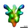
| Related structure data |  24708MC M: map data used to model this data C: citing same article ( |
|---|---|
| Similar structure data |


-Links
 PDBj
PDBj


- Assembly
| Deposited unit | 
|
|---|---|
| 1 |
|
-Components
| #1: Protein | Mass: 142399.375 Da / Num. of mol.: 3 Source method: isolated from a genetically manipulated source Source: (gene. exp.) Severe acute respiratory syndrome coronavirus 2Gene: S, 2 / Production host: Homo sapiens (human) / References: UniProt: P0DTC2#2: Antibody | Mass: 27018.072 Da / Num. of mol.: 3 Source method: isolated from a genetically manipulated source Source: (gene. exp.) Homo sapiens (human) / Production host: Homo sapiens (human)#3: Antibody | Mass: 23220.752 Da / Num. of mol.: 3 Source method: isolated from a genetically manipulated source Source: (gene. exp.) Homo sapiens (human) / Production host: Homo sapiens (human)#4: Polysaccharide | 2-acetamido-2-deoxy-beta-D-glucopyranose-(1-4)-2-acetamido-2-deoxy-beta-D-glucopyranose Source method: isolated from a genetically manipulated source #5: Sugar | ChemComp-NAG /   Type: D-saccharide, beta linking / Mass: 221.208 Da / Num. of mol.: 48 / Source method: obtained synthetically / Formula: C8H15NO6 / Feature type: SUBJECT OF INVESTIGATION Type: D-saccharide, beta linking / Mass: 221.208 Da / Num. of mol.: 48 / Source method: obtained synthetically / Formula: C8H15NO6 / Feature type: SUBJECT OF INVESTIGATIONHas ligand of interest | Y | Has protein modification | Y | |
|---|
-Experimental details
-Experiment
| Experiment | Method: ELECTRON MICROSCOPY |
|---|---|
| EM experiment | Aggregation state: PARTICLE / 3D reconstruction method: single particle reconstruction |
- Sample preparation
| Component | Name: Prefusion SARS-CoV-2 spike glycoprotein in complex with 5-7 Fab Type: COMPLEX / Entity ID: #1-#3 / Source: MULTIPLE SOURCES |
|---|---|
| Source (natural) | Organism: Severe acute respiratory syndrome coronavirus 2 |
| Source (recombinant) | Organism: Homo sapiens (human) |
| Buffer solution | pH: 4.5 |
| Specimen | Embedding applied: NO / Shadowing applied: NO / Staining applied: NO / Vitrification applied: YES |
| Vitrification | Cryogen name: ETHANE |
- Electron microscopy imaging
| Experimental equipment |  Model: Titan Krios / Image courtesy: FEI Company |
|---|---|
| Microscopy | Model: FEI TITAN KRIOS |
| Electron gun | Electron source:  FIELD EMISSION GUN / Accelerating voltage: 300 kV / Illumination mode: OTHER FIELD EMISSION GUN / Accelerating voltage: 300 kV / Illumination mode: OTHER |
| Electron lens | Mode: BRIGHT FIELD |
| Image recording | Electron dose: 42 e/Å2 / Film or detector model: GATAN K3 BIOQUANTUM (6k x 4k) |
- Processing
| Software | Name: PHENIX / Version: dev_4301: / Classification: refinement | ||||||||||||||||||||||||
|---|---|---|---|---|---|---|---|---|---|---|---|---|---|---|---|---|---|---|---|---|---|---|---|---|---|
| EM software |
| ||||||||||||||||||||||||
| CTF correction | Type: PHASE FLIPPING AND AMPLITUDE CORRECTION | ||||||||||||||||||||||||
| 3D reconstruction | Resolution: 3.5 Å / Resolution method: FSC 0.143 CUT-OFF / Num. of particles: 50866 / Symmetry type: POINT | ||||||||||||||||||||||||
| Atomic model building | Protocol: FLEXIBLE FIT | ||||||||||||||||||||||||
| Refine LS restraints |
|
