 Movie
Movie Controller

[English] 日本語
 Yorodumi
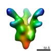
- PDB-6vsj: Cryo-electron microscopy structure of mouse coronavirus spike pro... -
+ Open data

- Basic information
| Entry | Database: PDB / ID: 6vsj | ||||||
|---|---|---|---|---|---|---|---|
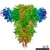
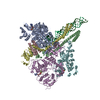
| Title | Cryo-electron microscopy structure of mouse coronavirus spike protein complexed with its murine receptor | ||||||
 Components Components |
| ||||||
 Keywords Keywords | VIRAL PROTEIN / MHV spike / CEACAM1a / Complex / Glycoprotein | ||||||
| Function / homology |  Function and homology information Function and homology informationgranulocyte colony-stimulating factor receptor binding / Fibronectin matrix formation / positive regulation of homophilic cell adhesion / Post-translational modification: synthesis of GPI-anchored proteins / negative regulation of cytotoxic T cell degranulation / regulation of endothelial cell differentiation / insulin receptor internalization / regulation of homophilic cell adhesion / granulocyte colony-stimulating factor signaling pathway / negative regulation of hepatocyte proliferation ...granulocyte colony-stimulating factor receptor binding / Fibronectin matrix formation / positive regulation of homophilic cell adhesion / Post-translational modification: synthesis of GPI-anchored proteins / negative regulation of cytotoxic T cell degranulation / regulation of endothelial cell differentiation / insulin receptor internalization / regulation of homophilic cell adhesion / granulocyte colony-stimulating factor signaling pathway / negative regulation of hepatocyte proliferation / regulation of epidermal growth factor receptor signaling pathway / regulation of blood vessel remodeling / regulation of sprouting angiogenesis / filamin binding / negative regulation of lipid biosynthetic process / negative regulation of natural killer cell mediated cytotoxicity directed against tumor cell target / negative regulation of T cell mediated cytotoxicity / regulation of endothelial cell migration / insulin catabolic process / Cell surface interactions at the vascular wall / common myeloid progenitor cell proliferation / negative regulation of granulocyte differentiation / Toll-like receptor binding / regulation of phosphatidylinositol 3-kinase/protein kinase B signal transduction / negative regulation of interleukin-1 production / : / positive regulation of vasculogenesis / negative regulation of fatty acid biosynthetic process / negative regulation of platelet aggregation / bile acid transmembrane transporter activity / cell-cell junction organization / regulation of immune system process / wound healing, spreading of cells / negative regulation of vascular permeability / negative regulation of bone resorption / negative regulation of JNK cascade / negative regulation of cytokine production / negative regulation of interleukin-2 production / ciliary membrane / bile acid and bile salt transport / blood vessel development / microvillus membrane / negative regulation of osteoclast differentiation / homophilic cell-cell adhesion / lateral plasma membrane / negative regulation of T cell receptor signaling pathway / regulation of ERK1 and ERK2 cascade / negative regulation of protein kinase activity / transport vesicle / negative regulation of T cell proliferation / Neutrophil degranulation / regulation of cell migration / basal plasma membrane / protein tyrosine kinase binding / adherens junction / regulation of cell growth / positive regulation of JNK cascade / kinase binding / cellular response to insulin stimulus / cell-cell junction / cell junction / virus receptor activity / actin binding / protein phosphatase binding / host cell Golgi apparatus / host cell endoplasmic reticulum-Golgi intermediate compartment membrane / receptor-mediated virion attachment to host cell / calmodulin binding / cell adhesion / protein dimerization activity / apical plasma membrane / endocytosis involved in viral entry into host cell / fusion of virus membrane with host plasma membrane / external side of plasma membrane / fusion of virus membrane with host endosome membrane / viral envelope / protein kinase binding / host cell plasma membrane / virion membrane / cell surface / signal transduction / protein homodimerization activity / membrane / identical protein binding / plasma membrane Similarity search - Function | ||||||
| Biological species |  Murine coronavirus Murine coronavirus | ||||||
| Method | ELECTRON MICROSCOPY / single particle reconstruction / cryo EM / Resolution: 3.94 Å | ||||||
 Authors Authors | Shang, J. / Wan, Y.S. / Liu, C. / Yount, B. / Gully, K. / Yang, Y. / Auerbach, A. / Peng, G.Q. / Baric, R. / Li, F. | ||||||
| Funding support |  United States, 1items United States, 1items
| ||||||
 Citation Citation | Journal: PLoS Pathog / Year: 2020 Title: Structure of mouse coronavirus spike protein complexed with receptor reveals mechanism for viral entry. Authors: Jian Shang / Yushun Wan / Chang Liu / Boyd Yount / Kendra Gully / Yang Yang / Ashley Auerbach / Guiqing Peng / Ralph Baric / Fang Li /  Abstract: Coronaviruses recognize a variety of receptors using different domains of their envelope-anchored spike protein. How these diverse receptor recognition patterns affect viral entry is unknown. Mouse ...Coronaviruses recognize a variety of receptors using different domains of their envelope-anchored spike protein. How these diverse receptor recognition patterns affect viral entry is unknown. Mouse hepatitis coronavirus (MHV) is the only known coronavirus that uses the N-terminal domain (NTD) of its spike to recognize a protein receptor, CEACAM1a. Here we determined the cryo-EM structure of MHV spike complexed with mouse CEACAM1a. The trimeric spike contains three receptor-binding S1 heads sitting on top of a trimeric membrane-fusion S2 stalk. Three receptor molecules bind to the sides of the spike trimer, where three NTDs are located. Receptor binding induces structural changes in the spike, weakening the interactions between S1 and S2. Using protease sensitivity and negative-stain EM analyses, we further showed that after protease treatment of the spike, receptor binding facilitated the dissociation of S1 from S2, allowing S2 to transition from pre-fusion to post-fusion conformation. Together these results reveal a new role of receptor binding in MHV entry: in addition to its well-characterized role in viral attachment to host cells, receptor binding also induces the conformational change of the spike and hence the fusion of viral and host membranes. Our study provides new mechanistic insight into coronavirus entry and highlights the diverse entry mechanisms used by different viruses. | ||||||
| History |
|
- Structure visualization
| Movie |
Movie viewer |
|---|---|
| Structure viewer | Molecule: MolmilJmol/JSmol |
- Downloads & links
-Download
| PDBx/mmCIF format | 6vsj.cif.gz | 653.8 KB | Display | PDBx/mmCIF format |
|---|---|---|---|---|
| PDB format | pdb6vsj.ent.gz | 523.4 KB | Display | PDB format |
| PDBx/mmJSON format | 6vsj.json.gz | Tree view | PDBx/mmJSON format | |
| Others |  Other downloads Other downloads |
-Validation report
| Arichive directory | https://data.pdbj.org/pub/pdb/validation_reports/vs/6vsjftp://data.pdbj.org/pub/pdb/validation_reports/vs/6vsj | HTTPS FTP |
|---|
-Related structure data
| Related structure data |  21377MC M: map data used to model this data C: citing same article ( |
|---|---|
| Similar structure data |


-Links
 PDBj
PDBj


- Assembly
| Deposited unit | 
|
|---|---|
| 1 |
|
-Components
| #1: Protein | Mass: 141118.797 Da / Num. of mol.: 3 Source method: isolated from a genetically manipulated source Source: (gene. exp.) Murine coronavirus (strain A59) / Strain: A59 / Gene: S, 3 / Production host: Baculovirus expression vector pFastBac1-HM / References: UniProt: P11224#2: Protein | Mass: 23678.691 Da / Num. of mol.: 3 Source method: isolated from a genetically manipulated source Source: (gene. exp.) Baculovirus expression vector pFastBac1-HM / References: UniProt: P31809, UniProt: Q3LFS9*PLUS#3: Sugar | ChemComp-NAG /   Type: D-saccharide, beta linking / Mass: 221.208 Da / Num. of mol.: 12 Type: D-saccharide, beta linking / Mass: 221.208 Da / Num. of mol.: 12Source method: isolated from a genetically manipulated source Formula: C8H15NO6 Has ligand of interest | N | Has protein modification | Y | |
|---|
-Experimental details
-Experiment
| Experiment | Method: ELECTRON MICROSCOPY |
|---|---|
| EM experiment | Aggregation state: PARTICLE / 3D reconstruction method: single particle reconstruction |
- Sample preparation
| Component |
| ||||||||||||||||||||||||
|---|---|---|---|---|---|---|---|---|---|---|---|---|---|---|---|---|---|---|---|---|---|---|---|---|---|
| Source (natural) |
| ||||||||||||||||||||||||
| Source (recombinant) |
| ||||||||||||||||||||||||
| Buffer solution | pH: 7.4 | ||||||||||||||||||||||||
| Buffer component |
| ||||||||||||||||||||||||
| Specimen | Conc.: 0.3 mg/ml / Embedding applied: NO / Shadowing applied: NO / Staining applied: NO / Vitrification applied: YES | ||||||||||||||||||||||||
| Vitrification | Cryogen name: ETHANE |
- Electron microscopy imaging
| Experimental equipment |  Model: Titan Krios / Image courtesy: FEI Company |
|---|---|
| Microscopy | Model: FEI TITAN KRIOS |
| Electron gun | Electron source:  FIELD EMISSION GUN / Accelerating voltage: 300 kV / Illumination mode: FLOOD BEAM FIELD EMISSION GUN / Accelerating voltage: 300 kV / Illumination mode: FLOOD BEAM |
| Electron lens | Mode: BRIGHT FIELD |
| Image recording | Electron dose: 77 e/Å2 / Detector mode: SUPER-RESOLUTION / Film or detector model: GATAN K2 SUMMIT (4k x 4k) |
- Processing
| CTF correction | Type: NONE |
|---|---|
| Symmetry | Point symmetry: C3 (3 fold cyclic) |
| 3D reconstruction | Resolution: 3.94 Å / Resolution method: FSC 0.143 CUT-OFF / Num. of particles: 82923 / Symmetry type: POINT |
