 Movie
Movie Controller

[English] 日本語
 Yorodumi
- PDB-7rut: Structure of Human ATP:Cobalamin Adenosyltransferase R190C bound ... -
+ Open data

- Basic information
| Entry | Database: PDB / ID: 7rut | ||||||
|---|---|---|---|---|---|---|---|
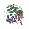
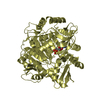
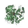
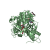
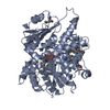
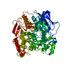
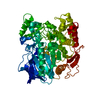
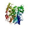
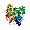
| Title | Structure of Human ATP:Cobalamin Adenosyltransferase R190C bound to ATP | ||||||
 Components Components | Corrinoid adenosyltransferase | ||||||
 Keywords Keywords | TRANSFERASE / ATP:cobalamin adenosyl transferase / cobalamin / vitamin B12 / patient mutation | ||||||
| Function / homology |  Function and homology information Function and homology informationDefective MMAB causes MMA, cblB type / Cobalamin (Cbl) metabolism / cobalamin metabolic process / corrinoid adenosyltransferase activity / Transferases; Transferring alkyl or aryl groups, other than methyl groups / transferase activity, transferring alkyl or aryl (other than methyl) groups / cobalamin binding / mitochondrial matrix / mitochondrion / ATP binding Similarity search - Function | ||||||
| Biological species |  Homo sapiens (human) Homo sapiens (human) | ||||||
| Method |  X-RAY DIFFRACTION / SYNCHROTRON / MOLECULAR REPLACEMENT / Resolution: 1.5 Å X-RAY DIFFRACTION / SYNCHROTRON / MOLECULAR REPLACEMENT / Resolution: 1.5 Å | ||||||
 Authors Authors | Mascarenhas, R. / Gouda, H. / Koutmos, M. / Banerjee, R. | ||||||
| Funding support |  United States, 1items United States, 1items
| ||||||
 Citation Citation | Journal: J.Biol.Chem. / Year: 2021 Title: Patient mutations in human ATP:cob(I)alamin adenosyltransferase differentially affect its catalytic versus chaperone functions. Authors: Gouda, H. / Mascarenhas, R. / Pillay, S. / Ruetz, M. / Koutmos, M. / Banerjee, R. | ||||||
| History |
|
- Structure visualization
| Structure viewer | Molecule: MolmilJmol/JSmol |
|---|
- Downloads & links
-Download
| PDBx/mmCIF format | 7rut.cif.gz | 470.8 KB | Display | PDBx/mmCIF format |
|---|---|---|---|---|
| PDB format | pdb7rut.ent.gz | 382.2 KB | Display | PDB format |
| PDBx/mmJSON format | 7rut.json.gz | Tree view | PDBx/mmJSON format | |
| Others |  Other downloads Other downloads |
-Validation report
| Arichive directory | https://data.pdbj.org/pub/pdb/validation_reports/ru/7rutftp://data.pdbj.org/pub/pdb/validation_reports/ru/7rut | HTTPS FTP |
|---|
-Related structure data
| Related structure data |  7ruuC  7ruvC  2idxS S: Starting model for refinement C: citing same article ( |
|---|---|
| Similar structure data |

-Links
 PDBj
PDBj- Assembly
| Deposited unit | 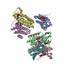
| ||||||||
|---|---|---|---|---|---|---|---|---|---|
| 1 | 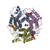
| ||||||||
| 2 | 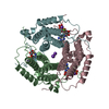
| ||||||||
| Unit cell |
|
-Components
-Protein , 1 types, 6 molecules ECFBAD
| #1: Protein | Mass: 21666.643 Da / Num. of mol.: 6 / Mutation: R190C Source method: isolated from a genetically manipulated source Source: (gene. exp.) Homo sapiens (human) / Gene: MMAB / Production host:  |
|---|
-Non-polymers , 5 types, 744 molecules 


| #2: Chemical | ChemComp-MG /  Mass: 24.305 Da / Num. of mol.: 9 / Source method: obtained synthetically / Formula: Mg Mass: 24.305 Da / Num. of mol.: 9 / Source method: obtained synthetically / Formula: Mg#3: Chemical | ChemComp-K /  Mass: 39.098 Da / Num. of mol.: 11 / Source method: obtained synthetically / Formula: K Mass: 39.098 Da / Num. of mol.: 11 / Source method: obtained synthetically / Formula: K#4: Chemical | ChemComp-ATP /  Mass: 507.181 Da / Num. of mol.: 6 / Source method: obtained synthetically / Formula: C10H16N5O13P3 / Feature type: SUBJECT OF INVESTIGATION / Comment: ATP, energy-carrying molecule*YM Mass: 507.181 Da / Num. of mol.: 6 / Source method: obtained synthetically / Formula: C10H16N5O13P3 / Feature type: SUBJECT OF INVESTIGATION / Comment: ATP, energy-carrying molecule*YM#5: Chemical | ChemComp-GOL / |  Mass: 92.094 Da / Num. of mol.: 1 / Source method: obtained synthetically / Formula: C3H8O3 Mass: 92.094 Da / Num. of mol.: 1 / Source method: obtained synthetically / Formula: C3H8O3#6: Water | ChemComp-HOH / | Mass: 18.015 Da / Num. of mol.: 717 / Source method: isolated from a natural source / Formula: H2O |
|---|
-Details
| Has ligand of interest | Y |
|---|
-Experimental details
-Experiment
| Experiment | Method: X-RAY DIFFRACTION / Number of used crystals: 1 |
|---|
- Sample preparation
| Crystal | Density Matthews: 2.15 Å3/Da / Density % sol: 42.91 % |
|---|---|
| Crystal grow | Temperature: 293 K / Method: vapor diffusion, hanging drop Details: 30% (v/v) 2-propanol, 100 mM Tris pH 8.5, and 30% (w/v) PEG 3350 |
-Data collection
| Diffraction | Mean temperature: 100 K / Serial crystal experiment: N |
|---|---|
| Diffraction source | Source: SYNCHROTRON / Site: APS / Beamline: 23-ID-B / Wavelength: 1.0332 Å |
| Detector | Type: DECTRIS EIGER X 16M / Detector: PIXEL / Date: Nov 15, 2019 |
| Radiation | Protocol: SINGLE WAVELENGTH / Monochromatic (M) / Laue (L): M / Scattering type: x-ray |
| Radiation wavelength | Wavelength: 1.0332 Å / Relative weight: 1 |
| Reflection | Resolution: 1.5→47.9 Å / Num. obs: 176322 / % possible obs: 99.9 % / Redundancy: 6.6 % / CC1/2: 1 / Rmerge(I) obs: 0.06 / Net I/σ(I): 12.8 |
| Reflection shell | Resolution: 1.5→1.53 Å / Rmerge(I) obs: 1.072 / Num. unique obs: 8753 / CC1/2: 0.66 |
- Processing
| Software |
| |||||||||||||||||||||||||||||||||||||||||||||||||||||||||||||||||||||||||||||||||||||||||||||||||||||||||||||||||||||||||||||||||||||||||||||||||||||||||||
|---|---|---|---|---|---|---|---|---|---|---|---|---|---|---|---|---|---|---|---|---|---|---|---|---|---|---|---|---|---|---|---|---|---|---|---|---|---|---|---|---|---|---|---|---|---|---|---|---|---|---|---|---|---|---|---|---|---|---|---|---|---|---|---|---|---|---|---|---|---|---|---|---|---|---|---|---|---|---|---|---|---|---|---|---|---|---|---|---|---|---|---|---|---|---|---|---|---|---|---|---|---|---|---|---|---|---|---|---|---|---|---|---|---|---|---|---|---|---|---|---|---|---|---|---|---|---|---|---|---|---|---|---|---|---|---|---|---|---|---|---|---|---|---|---|---|---|---|---|---|---|---|---|---|---|---|---|
| Refinement | Method to determine structure: MOLECULAR REPLACEMENT Starting model: 2IDX Resolution: 1.5→40.494 Å / SU ML: 0.16 / Cross valid method: THROUGHOUT / σ(F): 1.34 / Phase error: 19.29 / Stereochemistry target values: ML
| |||||||||||||||||||||||||||||||||||||||||||||||||||||||||||||||||||||||||||||||||||||||||||||||||||||||||||||||||||||||||||||||||||||||||||||||||||||||||||
| Solvent computation | Shrinkage radii: 0.9 Å / VDW probe radii: 1.11 Å / Solvent model: FLAT BULK SOLVENT MODEL | |||||||||||||||||||||||||||||||||||||||||||||||||||||||||||||||||||||||||||||||||||||||||||||||||||||||||||||||||||||||||||||||||||||||||||||||||||||||||||
| Displacement parameters | Biso max: 172.32 Å2 / Biso mean: 31.7175 Å2 / Biso min: 13.06 Å2 | |||||||||||||||||||||||||||||||||||||||||||||||||||||||||||||||||||||||||||||||||||||||||||||||||||||||||||||||||||||||||||||||||||||||||||||||||||||||||||
| Refinement step | Cycle: final / Resolution: 1.5→40.494 Å
| |||||||||||||||||||||||||||||||||||||||||||||||||||||||||||||||||||||||||||||||||||||||||||||||||||||||||||||||||||||||||||||||||||||||||||||||||||||||||||
| LS refinement shell | Refine-ID: X-RAY DIFFRACTION / Rfactor Rfree error: 0 / % reflection obs: 100 %
|
