 Movie
Movie Controller


+ Open data

- Basic information
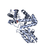
| Entry | Database: PDB / ID: 7rum | ||||||
|---|---|---|---|---|---|---|---|
| Title | Endolysin from Escherichia coli O157:H7 phage FTEbC1, LysT84 | ||||||
 Components Components | Endolysin | ||||||
 Keywords Keywords | HYDROLASE / Endolysin / peptidoglycan hydrolase / bacteriophage | ||||||
| Function / homology | N-acetylmuramidase / N-acetylmuramidase / PGBD superfamily / Peptidoglycan binding-like / Putative peptidoglycan binding domain / PGBD-like superfamily / Endolysin Function and homology information Function and homology information | ||||||
| Biological species |  Salmonella phage GEC_vB_GOT (virus) Salmonella phage GEC_vB_GOT (virus) | ||||||
| Method |  X-RAY DIFFRACTION / SYNCHROTRON / MOLECULAR REPLACEMENT / Resolution: 2.99 Å X-RAY DIFFRACTION / SYNCHROTRON / MOLECULAR REPLACEMENT / Resolution: 2.99 Å | ||||||
 Authors Authors | Love, M.J. / Billington, C. / Dobson, R.C.J. | ||||||
| Funding support | 1items
| ||||||
 Citation Citation | Journal: Biochem.J. / Year: 2022 Title: The structure and function of modular Escherichia coli O157:H7 bacteriophage FTBEc1 endolysin, LysT84: defining a new endolysin catalytic subfamily. Authors: Love, M.J. / Coombes, D. / Ismail, S. / Billington, C. / Dobson, R.C.J. | ||||||
| History |
|
- Structure visualization
| Structure viewer | Molecule: MolmilJmol/JSmol |
|---|
- Downloads & links
-Download
| PDBx/mmCIF format | 7rum.cif.gz | 256.4 KB | Display | PDBx/mmCIF format |
|---|---|---|---|---|
| PDB format | pdb7rum.ent.gz | 173 KB | Display | PDB format |
| PDBx/mmJSON format | 7rum.json.gz | Tree view | PDBx/mmJSON format | |
| Others |  Other downloads Other downloads |
-Validation report
| Arichive directory | https://data.pdbj.org/pub/pdb/validation_reports/ru/7rumftp://data.pdbj.org/pub/pdb/validation_reports/ru/7rum | HTTPS FTP |
|---|
-Related structure data
| Related structure data |  5nm7S S: Starting model for refinement |
|---|---|
| Similar structure data |


-Links
 PDBj
PDBj
- Assembly
| Deposited unit | 
| ||||||||||||||||||||||||||||||||||||||||||||||||||||||||||
|---|---|---|---|---|---|---|---|---|---|---|---|---|---|---|---|---|---|---|---|---|---|---|---|---|---|---|---|---|---|---|---|---|---|---|---|---|---|---|---|---|---|---|---|---|---|---|---|---|---|---|---|---|---|---|---|---|---|---|---|
| 1 | 
| ||||||||||||||||||||||||||||||||||||||||||||||||||||||||||
| 2 | 
| ||||||||||||||||||||||||||||||||||||||||||||||||||||||||||
| Unit cell |
| ||||||||||||||||||||||||||||||||||||||||||||||||||||||||||
| Components on special symmetry positions |
| ||||||||||||||||||||||||||||||||||||||||||||||||||||||||||
| Noncrystallographic symmetry (NCS) | NCS domain:
NCS domain segments:
NCS oper: (Code: givenMatrix: (0.872399463877, -0.488645226443, -0.0120423461758), (-0.488789308071, -0.872230163196, -0.0173076493567), (-0.00204639733111, 0.0209853540746, -0.999777688875)Vector: - ...NCS oper: (Code: given Matrix: (0.872399463877, -0.488645226443, -0.0120423461758), Vector: |
-Components
| #1: Protein | Mass: 31257.457 Da / Num. of mol.: 2 Source method: isolated from a genetically manipulated source Source: (gene. exp.) Salmonella phage GEC_vB_GOT (virus) / Gene: GECvBGOT_gp079c / Production host:  #2: Chemical | ChemComp-GOL /   Mass: 92.094 Da / Num. of mol.: 4 / Source method: obtained synthetically / Formula: C3H8O3 Mass: 92.094 Da / Num. of mol.: 4 / Source method: obtained synthetically / Formula: C3H8O3#3: Water | ChemComp-HOH / |  Mass: 18.015 Da / Num. of mol.: 87 / Source method: isolated from a natural source / Formula: H2O Mass: 18.015 Da / Num. of mol.: 87 / Source method: isolated from a natural source / Formula: H2OHas ligand of interest | N | Has protein modification | Y | |
|---|
-Experimental details
-Experiment
| Experiment | Method: X-RAY DIFFRACTION / Number of used crystals: 1 |
|---|
- Sample preparation
| Crystal | Density Matthews: 5.32 Å3/Da / Density % sol: 76.89 % |
|---|---|
| Crystal grow | Temperature: 293.15 K / Method: vapor diffusion, sitting drop Details: Morpheus I condition F3: 0.12 M monosaccharides [0.2 M D-glucose; 0.2 M D-mannose; 0.2 M D-galactose; 0.2 M L-fucose; 0.2 M D-xylose; 0.2 M N-acetyl-D-glucosamine] 0.1 M buffer System 1 [1.0 ...Details: Morpheus I condition F3: 0.12 M monosaccharides [0.2 M D-glucose; 0.2 M D-mannose; 0.2 M D-galactose; 0.2 M L-fucose; 0.2 M D-xylose; 0.2 M N-acetyl-D-glucosamine] 0.1 M buffer System 1 [1.0 M imidazole; MES monohydrate (acid) pH 6.5], 30% v/v Precipitant Mix 3 [40% v/v glycerol; 20% w/v PEG 4000 |
-Data collection
| Diffraction | Mean temperature: 110 K / Serial crystal experiment: N | |||||||||||||||||||||||||||
|---|---|---|---|---|---|---|---|---|---|---|---|---|---|---|---|---|---|---|---|---|---|---|---|---|---|---|---|---|
| Diffraction source | Source: SYNCHROTRON / Site: Australian Synchrotron  / Beamline: MX2 / Wavelength: 0.9537 Å / Beamline: MX2 / Wavelength: 0.9537 Å | |||||||||||||||||||||||||||
| Detector | Type: DECTRIS EIGER X 16M / Detector: PIXEL / Date: Mar 19, 2019 | |||||||||||||||||||||||||||
| Radiation | Protocol: SINGLE WAVELENGTH / Monochromatic (M) / Laue (L): M / Scattering type: x-ray | |||||||||||||||||||||||||||
| Radiation wavelength | Wavelength: 0.9537 Å / Relative weight: 1 | |||||||||||||||||||||||||||
| Reflection | Resolution: 2.99→49.04 Å / Num. obs: 26401 / % possible obs: 99.9 % / Redundancy: 18.5 % / CC1/2: 0.974 / Rmerge(I) obs: 0.521 / Rpim(I) all: 0.124 / Rrim(I) all: 0.535 / Net I/σ(I): 6.6 / Num. measured all: 487418 / Scaling rejects: 92 | |||||||||||||||||||||||||||
| Reflection shell | Diffraction-ID: 1 / % possible all: 99.5
|
- Processing
| Software |
| |||||||||||||||||||||||||||||||||||||||||||||||||||||||||||||||||||||||||||||
|---|---|---|---|---|---|---|---|---|---|---|---|---|---|---|---|---|---|---|---|---|---|---|---|---|---|---|---|---|---|---|---|---|---|---|---|---|---|---|---|---|---|---|---|---|---|---|---|---|---|---|---|---|---|---|---|---|---|---|---|---|---|---|---|---|---|---|---|---|---|---|---|---|---|---|---|---|---|---|
| Refinement | Method to determine structure: MOLECULAR REPLACEMENT Starting model: 5NM7 Resolution: 2.99→49.04 Å / SU ML: 0.3929 / Cross valid method: FREE R-VALUE / σ(F): 1.92 / Phase error: 25.8838 Stereochemistry target values: GeoStd + Monomer Library + CDL v1.2
| |||||||||||||||||||||||||||||||||||||||||||||||||||||||||||||||||||||||||||||
| Solvent computation | Shrinkage radii: 0.9 Å / VDW probe radii: 1.11 Å / Solvent model: FLAT BULK SOLVENT MODEL | |||||||||||||||||||||||||||||||||||||||||||||||||||||||||||||||||||||||||||||
| Displacement parameters | Biso mean: 62.27 Å2 | |||||||||||||||||||||||||||||||||||||||||||||||||||||||||||||||||||||||||||||
| Refinement step | Cycle: LAST / Resolution: 2.99→49.04 Å
| |||||||||||||||||||||||||||||||||||||||||||||||||||||||||||||||||||||||||||||
| Refine LS restraints |
| |||||||||||||||||||||||||||||||||||||||||||||||||||||||||||||||||||||||||||||
| Refine LS restraints NCS | Type: Torsion NCS / Rms dev position: 0.837109117449 Å | |||||||||||||||||||||||||||||||||||||||||||||||||||||||||||||||||||||||||||||
| LS refinement shell |
| |||||||||||||||||||||||||||||||||||||||||||||||||||||||||||||||||||||||||||||
| Refinement TLS params. | Method: refined / Origin x: -23.3299402913 Å / Origin y: -22.4033130523 Å / Origin z: 15.3999379554 Å
| |||||||||||||||||||||||||||||||||||||||||||||||||||||||||||||||||||||||||||||
| Refinement TLS group | Selection details: all |
