 Movie
Movie Controller

[English] 日本語
 Yorodumi
- PDB-7rr4: Structure of Deep-Sea Phage NrS-1 Primase-Polymerase N300 in comp... -
+ Open data

- Basic information
| Entry | Database: PDB / ID: 7rr4 | ||||||
|---|---|---|---|---|---|---|---|
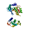
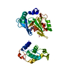
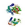
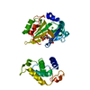
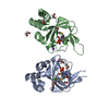
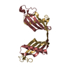
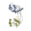
| Title | Structure of Deep-Sea Phage NrS-1 Primase-Polymerase N300 in complex with magnesium and pyrophosphate | ||||||
 Components Components | Primase | ||||||
 Keywords Keywords | REPLICATION / primpol / denovo / polymerase / nrspol | ||||||
| Function / homology |  Function and homology information Function and homology informationviral DNA genome replication / DNA helicase activity / Transferases; Transferring phosphorus-containing groups; Nucleotidyltransferases / DNA-directed DNA polymerase / DNA helicase / DNA-directed DNA polymerase activity / DNA replication / hydrolase activity / ATP binding Similarity search - Function | ||||||
| Biological species |  Nitratiruptor phage NrS-1 (virus) Nitratiruptor phage NrS-1 (virus) | ||||||
| Method |  X-RAY DIFFRACTION / SYNCHROTRON / MOLECULAR REPLACEMENT / Resolution: 1.86 Å X-RAY DIFFRACTION / SYNCHROTRON / MOLECULAR REPLACEMENT / Resolution: 1.86 Å | ||||||
 Authors Authors | Wang, L. / Yu, C. / Sliz, P. | ||||||
| Funding support | 1items
| ||||||
 Citation Citation | Journal: Frontiers in Microbiology / Year: 2021 Title: Molecular Dissection of the Primase and Polymerase Activities of Deep-Sea Phage NrS-1 Primase-Polymerase Authors: Huang, F. / Lu, X. / Yu, C. / Sliz, P. / Wang, L. / Zhu, B. | ||||||
| History |
|
- Structure visualization
| Structure viewer | Molecule: MolmilJmol/JSmol |
|---|
- Downloads & links
-Download
| PDBx/mmCIF format | 7rr4.cif.gz | 84.9 KB | Display | PDBx/mmCIF format |
|---|---|---|---|---|
| PDB format | pdb7rr4.ent.gz | 50.1 KB | Display | PDB format |
| PDBx/mmJSON format | 7rr4.json.gz | Tree view | PDBx/mmJSON format | |
| Others |  Other downloads Other downloads |
-Validation report
| Arichive directory | https://data.pdbj.org/pub/pdb/validation_reports/rr/7rr4ftp://data.pdbj.org/pub/pdb/validation_reports/rr/7rr4 | HTTPS FTP |
|---|
-Related structure data
| Related structure data |  7rr3S S: Starting model for refinement |
|---|---|
| Similar structure data |


-Links
 PDBj
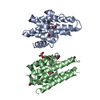
PDBj- Assembly
| Deposited unit | 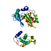
| ||||||||||||
|---|---|---|---|---|---|---|---|---|---|---|---|---|---|
| 1 |
| ||||||||||||
| Unit cell |
|
-Components
| #1: Protein | Mass: 32549.818 Da / Num. of mol.: 1 Source method: isolated from a genetically manipulated source Source: (gene. exp.) Nitratiruptor phage NrS-1 (virus) / Production host:  | ||||||
|---|---|---|---|---|---|---|---|
| #2: Chemical |   Mass: 24.305 Da / Num. of mol.: 2 / Source method: obtained synthetically / Formula: Mg Mass: 24.305 Da / Num. of mol.: 2 / Source method: obtained synthetically / Formula: Mg#3: Chemical | ChemComp-PPV / | 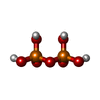  Mass: 177.975 Da / Num. of mol.: 1 / Source method: obtained synthetically / Formula: H4O7P2 / Feature type: SUBJECT OF INVESTIGATION Mass: 177.975 Da / Num. of mol.: 1 / Source method: obtained synthetically / Formula: H4O7P2 / Feature type: SUBJECT OF INVESTIGATION#4: Water | ChemComp-HOH / |  Mass: 18.015 Da / Num. of mol.: 100 / Source method: isolated from a natural source / Formula: H2O Mass: 18.015 Da / Num. of mol.: 100 / Source method: isolated from a natural source / Formula: H2OHas ligand of interest | Y | |
-Experimental details
-Experiment
| Experiment | Method: X-RAY DIFFRACTION / Number of used crystals: 1 |
|---|
- Sample preparation
| Crystal | Density Matthews: 2.66 Å3/Da / Density % sol: 53.71 % |
|---|---|
| Crystal grow | Temperature: 298 K / Method: vapor diffusion, sitting drop / pH: 4.5 Details: 800 mM sodium phosphate monobasic, 1200 mM potassium phosphate dibasic, and 100 mM sodium acetate (pH 4.5) |
-Data collection
| Diffraction | Mean temperature: 100 K / Serial crystal experiment: N |
|---|---|
| Diffraction source | Source: SYNCHROTRON / Site: APS  / Beamline: 24-ID-E / Wavelength: 0.979 Å / Beamline: 24-ID-E / Wavelength: 0.979 Å |
| Detector | Type: DECTRIS EIGER2 X 16M / Detector: PIXEL / Date: Oct 1, 2015 |
| Radiation | Protocol: SINGLE WAVELENGTH / Monochromatic (M) / Laue (L): M / Scattering type: x-ray |
| Radiation wavelength | Wavelength: 0.979 Å / Relative weight: 1 |
| Reflection | Resolution: 1.86→51.34 Å / Num. obs: 26979 / % possible obs: 98.29 % / Redundancy: 3.4 % / Biso Wilson estimate: 38.66 Å2 / Rmerge(I) obs: 0.09634 / Net I/σ(I): 12.8 |
| Reflection shell | Resolution: 1.86→1.93 Å / Rmerge(I) obs: 1.175 / Num. unique obs: 2708 |
- Processing
| Software |
| |||||||||||||||||||||||||||||||||||||||||||||||||||||||||||||||||||||||||||||
|---|---|---|---|---|---|---|---|---|---|---|---|---|---|---|---|---|---|---|---|---|---|---|---|---|---|---|---|---|---|---|---|---|---|---|---|---|---|---|---|---|---|---|---|---|---|---|---|---|---|---|---|---|---|---|---|---|---|---|---|---|---|---|---|---|---|---|---|---|---|---|---|---|---|---|---|---|---|---|
| Refinement | Method to determine structure: MOLECULAR REPLACEMENT Starting model: 7RR3 Resolution: 1.86→51.34 Å / SU ML: 0.2667 / Cross valid method: FREE R-VALUE / σ(F): 1.35 / Phase error: 29.6638 Stereochemistry target values: GeoStd + Monomer Library + CDL v1.2
| |||||||||||||||||||||||||||||||||||||||||||||||||||||||||||||||||||||||||||||
| Solvent computation | Shrinkage radii: 0.9 Å / VDW probe radii: 1.11 Å / Solvent model: FLAT BULK SOLVENT MODEL | |||||||||||||||||||||||||||||||||||||||||||||||||||||||||||||||||||||||||||||
| Displacement parameters | Biso mean: 48.09 Å2 | |||||||||||||||||||||||||||||||||||||||||||||||||||||||||||||||||||||||||||||
| Refinement step | Cycle: LAST / Resolution: 1.86→51.34 Å
| |||||||||||||||||||||||||||||||||||||||||||||||||||||||||||||||||||||||||||||
| Refine LS restraints |
| |||||||||||||||||||||||||||||||||||||||||||||||||||||||||||||||||||||||||||||
| LS refinement shell |
|
