[English] 日本語
Yorodumi
- PDB-3u34: Crystal structure of the general stress FMN/FAD binding protein f... -

+
Open data


ID or keywords:

Loading...

-
Basic information

Entry
Database: PDB / ID: 3u34
Title
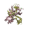
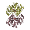
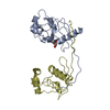
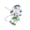
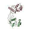
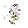
Crystal structure of the general stress FMN/FAD binding protein from the phytopathogen Xanthomonas citri
ComponentsGeneral stress protein
KeywordsPROTEIN BINDING / Xanthomonas citri General stress protein FMN binding protein FAD binding protein / PNP-oxidase like fold / FMN FAD
Function / homologyGeneral stress protein, FMN-binding split barrel domain / Pyridoxamine 5'-phosphate oxidase like / Electron Transport, Fmn-binding Protein; Chain A / Pnp Oxidase; Chain A / FMN-binding split barrel / Roll / Mainly Beta / TRIETHYLENE GLYCOL / General stress protein
Function and homology information
Biological speciesXanthomonas axonopodis pv. citri (bacteria)
MethodX-RAY DIFFRACTION / MOLECULAR REPLACEMENT / Resolution: 2.8 Å
AuthorsHilario, E. / Li, Y. / Fan, L.
CitationJournal: Acta Crystallogr.,Sect.D / Year: 2012
Title: The structure of a Xanthomonas general stress protein involved in citrus canker reveals its flavin-binding property.
Authors: Hilario, E. / Li, Y. / Niks, D. / Fan, L.
History
DepositionOct 4, 2011Deposition site: RCSB / Processing site: RCSB
Revision 1.0Jun 27, 2012Provider: repository / Type: Initial release
Revision 1.1Jan 23, 2013Group: Database references
Revision 1.2Oct 25, 2017Group: Author supporting evidence / Category: pdbx_struct_assembly_auth_evidence
Revision 1.3Sep 13, 2023Group: Data collection / Database references ...Data collection / Database references / Derived calculations / Refinement description
Category: chem_comp_atom / chem_comp_bond ...chem_comp_atom / chem_comp_bond / database_2 / pdbx_initial_refinement_model / struct_site
Item: _database_2.pdbx_DOI / _database_2.pdbx_database_accession ..._database_2.pdbx_DOI / _database_2.pdbx_database_accession / _struct_site.pdbx_auth_asym_id / _struct_site.pdbx_auth_comp_id / _struct_site.pdbx_auth_seq_id

-
Structure visualization

Structure viewerMolecule:
MolmilJmol/JSmol

Downloads & links

-
Assembly

Deposited unit
A: General stress protein
B: General stress protein
C: General stress protein
D: General stress protein
hetero molecules


Theoretical massNumber of molelcules
Total (without water)80,7366
Polymers80,4364
Non-polymers3002
Water84747
1


  • Idetical with deposited unit
  • defined by author&software
  • Evidence: gel filtration
TypeNameSymmetry operationNumber
identity operation1_555x,y,z1
Buried area11630 Å2
ΔGint-50 kcal/mol
Surface area24610 Å2
MethodPISA
2
A: General stress protein
D: General stress protein
hetero molecules


Theoretical massNumber of molelcules
Total (without water)40,3683
Polymers40,2182
Non-polymers1501
Water362
TypeNameSymmetry operationNumber
identity operation1_555x,y,z1
Buried area2880 Å2
ΔGint-14 kcal/mol
Surface area15250 Å2
MethodPISA
3
A: General stress protein
B: General stress protein
hetero molecules


Theoretical massNumber of molelcules
Total (without water)40,3683
Polymers40,2182
Non-polymers1501
Water362
TypeNameSymmetry operationNumber
identity operation1_555x,y,z1
Buried area2420 Å2
ΔGint-7 kcal/mol
Surface area15810 Å2
MethodPISA
4
B: General stress protein
C: General stress protein
hetero molecules


Theoretical massNumber of molelcules
Total (without water)40,3683
Polymers40,2182
Non-polymers1501
Water362
TypeNameSymmetry operationNumber
identity operation1_555x,y,z1
Buried area2740 Å2
ΔGint-15 kcal/mol
Surface area15360 Å2
MethodPISA
5
C: General stress protein
D: General stress protein
hetero molecules


Theoretical massNumber of molelcules
Total (without water)40,3683
Polymers40,2182
Non-polymers1501
Water362
TypeNameSymmetry operationNumber
identity operation1_555x,y,z1
Buried area2430 Å2
ΔGint-8 kcal/mol
Surface area15580 Å2
MethodPISA
Unit cell
Length a, b, c (Å)120.308, 122.135, 57.734
Angle α, β, γ (deg.)90.00, 90.00, 90.00
Int Tables number18
Space group name H-MP21212
Noncrystallographic symmetry (NCS)NCS domain:
IDEns-ID
11
21
31
41

NCS domain segments:
Dom-IDComponent-IDEns-IDSelection details
111chain A and (resseq 24:152 )
211chain B and (resseq 24:152 )
311chain C and (resseq 24:152 )
411chain D and (resseq 24:152 )
Details
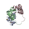
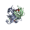
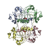
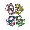
BASED ON THE SIZE EXCLUSION CHROMATOGRAPHY RESULTS THE PURE RECOMBINANT PROTEIN EXIST AS AN EQUILIBRIUM OF DIMERS AND TETRAMERS IN SOLUTION. HOWEVER, THE AMOUNT OF DIMERIC FORM IS PREDOMINANT WHEN COMPARED WITH THE TETRAMERIC FORM

-
Components

#1: Protein
General stress protein


Mass: 20108.908 Da / Num. of mol.: 4
Source method: isolated from a genetically manipulated source
Details: Gene was cloned into EcoRI and HindIII restriction sites of pET28a.
Source: (gene. exp.) Xanthomonas axonopodis pv. citri (bacteria)
Strain: 306 / Gene: General stress protein NCBI-GeneID 1156440, XAC2369 / Plasmid: pET28a / Production host: Escherichia coli (E. coli) / Strain (production host): BL21(DE3)pLys-S / References: UniProt: Q8PK08
#2: Chemical ChemComp-PGE / TRIETHYLENE GLYCOL


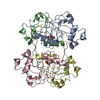
Mass: 150.173 Da / Num. of mol.: 2 / Source method: obtained synthetically / Formula: C6H14O4
#3: Water ChemComp-HOH / water


Mass: 18.015 Da / Num. of mol.: 47 / Source method: isolated from a natural source / Formula: H2O

-
Experimental details

-
Experiment

ExperimentMethod: X-RAY DIFFRACTION / Number of used crystals: 1

-
Sample preparation

CrystalDensity Matthews: 2.64 Å3/Da / Density % sol: 53.35 %
Crystal growTemperature: 298 K / Method: vapor diffusion, sitting drop / pH: 7.5
Details: Protein concentration at 25 mg/mL in 0.1M Hepes buffer, pH 7.5, containing 42% PEG200, VAPOR DIFFUSION, SITTING DROP, temperature 298K

-
Data collection

DiffractionMean temperature: 100 K
Diffraction sourceSource: ROTATING ANODE / Type: RIGAKU MICROMAX-007 HF / Wavelength: 1.54187 Å
DetectorType: RIGAKU RAXIS IV++ / Detector: IMAGE PLATE / Date: Feb 3, 2010 / Details: mirrors
RadiationMonochromator: mirror / Protocol: SINGLE WAVELENGTH / Monochromatic (M) / Laue (L): M / Scattering type: x-ray
Radiation wavelengthWavelength: 1.54187 Å / Relative weight: 1
ReflectionResolution: 2.8→20 Å / Num. all: 21644 / Num. obs: 20973 / % possible obs: 96.9 % / Observed criterion σ(F): 2 / Observed criterion σ(I): 2
Reflection shellResolution: 2.8→2.9 Å / % possible all: 95.7

-
Processing

Software
NameVersionClassification
CrystalClear2.0 r2data collection
PHASERphasing
PHENIX(phenix.refine: 1.6.1_357)refinement
HKL-2000data reduction
HKL-2000data scaling
RefinementMethod to determine structure: MOLECULAR REPLACEMENT
Starting model: 3DMB
Resolution: 2.8→19.807 Å / SU ML: 0.38 / σ(F): 0.05 / Phase error: 24.99 / Stereochemistry target values: ML
RfactorNum. reflection% reflectionSelection details
Rfree0.2602 1029 5.13 %RANDOM
Rwork0.2123 ---
obs0.2147 20050 92.71 %-
all-21627 --
Solvent computationShrinkage radii: 0.9 Å / VDW probe radii: 1.11 Å / Solvent model: FLAT BULK SOLVENT MODEL / Bsol: 40.539 Å2 / ksol: 0.339 e/Å3
Displacement parameters
Baniso -1Baniso -2Baniso -3
1-20.6129 Å2-0 Å2-0 Å2
2---11.4578 Å20 Å2
3----9.1551 Å2
Refinement stepCycle: LAST / Resolution: 2.8→19.807 Å
ProteinNucleic acidLigandSolventTotal
Num. atoms4263 0 20 47 4330
Refine LS restraints
Refine-IDTypeDev idealNumber
X-RAY DIFFRACTIONf_bond_d0.0124377
X-RAY DIFFRACTIONf_angle_d1.3145929
X-RAY DIFFRACTIONf_dihedral_angle_d15.9641539
X-RAY DIFFRACTIONf_chiral_restr0.088648
X-RAY DIFFRACTIONf_plane_restr0.005762
Refine LS restraints NCS
Ens-IDDom-IDAuth asym-IDNumberRefine-IDTypeRms dev position (Å)
11A973X-RAY DIFFRACTIONPOSITIONAL
12B973X-RAY DIFFRACTIONPOSITIONAL0.135
13C983X-RAY DIFFRACTIONPOSITIONAL0.194
14D979X-RAY DIFFRACTIONPOSITIONAL0.161
LS refinement shell
Resolution (Å)Rfactor RfreeNum. reflection RfreeRfactor RworkNum. reflection RworkRefine-ID% reflection obs (%)
2.7979-2.94490.37031390.27512378X-RAY DIFFRACTION83
2.9449-3.12880.32531350.23242515X-RAY DIFFRACTION87
3.1288-3.36920.27231690.22222673X-RAY DIFFRACTION93
3.3692-3.70630.27091530.19912810X-RAY DIFFRACTION97
3.7063-4.2380.24241330.18982866X-RAY DIFFRACTION98
4.238-5.32230.20691280.17712876X-RAY DIFFRACTION96
5.3223-19.80730.23691720.21982903X-RAY DIFFRACTION95
Refinement TLS params.

Method: refined / Refine-ID: X-RAY DIFFRACTION

IDL112)L122)L132)L222)L232)L332)S11 (Å °)S12 (Å °)S13 (Å °)S21 (Å °)S22 (Å °)S23 (Å °)S31 (Å °)S32 (Å °)S33 (Å °)T112)T122)T132)T222)T232)T332)Origin x (Å)Origin y (Å)Origin z (Å)
10.69530.3489-0.34821.1178-0.50281.00020.0969-0.12510.05440.2215-0.03070.0002-0.10340.0537-0.07820.2360.07050.0420.1154-0.0340.067-35.9746.37586.6879
20.7172-0.1194-0.15240.98450.66512.5696-0.05370.0592-0.1095-0.20660.1035-0.00650.6830.0551-0.07590.3856-0.01810.05850.1796-0.02340.0603-33.10425.6795-14.7724
30.6607-0.11540.5141.7480.20680.6223-0.10080.01360.0460.19560.0652-0.39670.0468-0.00320.01890.05530.0515-0.0170.0846-0.01890.1944-21.326631.9641.7165
40.6274-0.4112-0.21072.1804-0.3620.170.08790.27920.0185-0.1554-0.08870.56130.0429-0.0422-0.01550.0707-0.0193-0.03460.16490.00380.272-38.803934.0239-11.323
Refinement TLS group
IDRefine-IDRefine TLS-IDSelection details
1X-RAY DIFFRACTION1chain A
2X-RAY DIFFRACTION2chain B
3X-RAY DIFFRACTION3chain C
4X-RAY DIFFRACTION4chain D

+
About Yorodumi

-
News

-
Feb 9, 2022. New format data for meta-information of EMDB entries

New format data for meta-information of EMDB entries

  • Version 3 of the EMDB header file is now the official format.
  • The previous official version 1.9 will be removed from the archive.

Related info.:EMDB header

External links:wwPDB to switch to version 3 of the EMDB data model

-
Aug 12, 2020. Covid-19 info

Covid-19 info

URL: https://pdbjlvh1.pdbj.org/emnavi/covid19.php

New page: Covid-19 featured information page in EM Navigator.

Related info.:Covid-19 info / Mar 5, 2020. Novel coronavirus structure data

+
Mar 5, 2020. Novel coronavirus structure data

Novel coronavirus structure data

Related info.:Yorodumi Speices / Aug 12, 2020. Covid-19 info

External links:COVID-19 featured content - PDBj / Molecule of the Month (242):Coronavirus Proteases

+
Jan 31, 2019. EMDB accession codes are about to change! (news from PDBe EMDB page)

EMDB accession codes are about to change! (news from PDBe EMDB page)

  • The allocation of 4 digits for EMDB accession codes will soon come to an end. Whilst these codes will remain in use, new EMDB accession codes will include an additional digit and will expand incrementally as the available range of codes is exhausted. The current 4-digit format prefixed with “EMD-” (i.e. EMD-XXXX) will advance to a 5-digit format (i.e. EMD-XXXXX), and so on. It is currently estimated that the 4-digit codes will be depleted around Spring 2019, at which point the 5-digit format will come into force.
  • The EM Navigator/Yorodumi systems omit the EMD- prefix.

Related info.:Q: What is EMD? / ID/Accession-code notation in Yorodumi/EM Navigator

External links:EMDB Accession Codes are Changing Soon! / Contact to PDBj

+
Jul 12, 2017. Major update of PDB

Major update of PDB

  • wwPDB released updated PDB data conforming to the new PDBx/mmCIF dictionary.
  • This is a major update changing the version number from 4 to 5, and with Remediation, in which all the entries are updated.
  • In this update, many items about electron microscopy experimental information are reorganized (e.g. em_software).
  • Now, EM Navigator and Yorodumi are based on the updated data.

External links:wwPDB Remediation / Enriched Model Files Conforming to OneDep Data Standards Now Available in the PDB FTP Archive

-
Yorodumi

Thousand views of thousand structures

  • Yorodumi is a browser for structure data from EMDB, PDB, SASBDB, etc.
  • This page is also the successor to EM Navigator detail page, and also detail information page/front-end page for Omokage search.
  • The word "yorodu" (or yorozu) is an old Japanese word meaning "ten thousand". "mi" (miru) is to see.

Related info.:EMDB / PDB / SASBDB / Comparison of 3 databanks / Yorodumi Search / Aug 31, 2016. New EM Navigator & Yorodumi / Yorodumi Papers / Jmol/JSmol / Function and homology information / Changes in new EM Navigator and Yorodumi

Read more