 Movie
Movie Controller

[English] 日本語
 Yorodumi
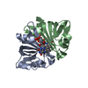
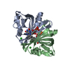
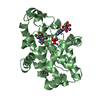
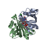
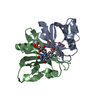
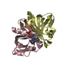
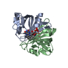
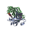
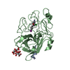
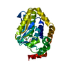
- PDB-1flm: DIMER OF FMN-BINDING PROTEIN FROM DESULFOVIBRIO VULGARIS (MIYAZAKI F) -
+ Open data

- Basic information
| Entry | Database: PDB / ID: 1flm | ||||||
|---|---|---|---|---|---|---|---|
| Title | DIMER OF FMN-BINDING PROTEIN FROM DESULFOVIBRIO VULGARIS (MIYAZAKI F) | ||||||
 Components Components | PROTEIN (FMN-BINDING PROTEIN) | ||||||
 Keywords Keywords | ELECTRON TRANSPORT / FMN BINDING | ||||||
| Function / homology |  Function and homology information Function and homology information | ||||||
| Biological species |  Desulfovibrio vulgaris str. 'Miyazaki F' (bacteria) Desulfovibrio vulgaris str. 'Miyazaki F' (bacteria) | ||||||
| Method |  X-RAY DIFFRACTION / SYNCHROTRON / MIR / Resolution: 1.3 Å X-RAY DIFFRACTION / SYNCHROTRON / MIR / Resolution: 1.3 Å | ||||||
 Authors Authors | Suto, K. / Kawagoe, K. / Shibata, N. / Morimoto, K. / Higuchi, Y. / Kitamura, M. / Nakaya, T. / Yasuoka, N. | ||||||
 Citation Citation | Journal: Acta Crystallogr.,Sect.D / Year: 2000 Title: How do the x-ray structure and the NMR structure of FMN-binding protein differ? Authors: Suto, K. / Kawagoe, K. / Shibata, N. / Morimoto, Y. / Higuchi, Y. / Kitamura, M. / Nakaya, T. / Yasuoka, N. | ||||||
| History |
|
- Structure visualization
| Structure viewer | Molecule: MolmilJmol/JSmol |
|---|
- Downloads & links
-Download
| PDBx/mmCIF format | 1flm.cif.gz | 63.5 KB | Display | PDBx/mmCIF format |
|---|---|---|---|---|
| PDB format | pdb1flm.ent.gz | 46.3 KB | Display | PDB format |
| PDBx/mmJSON format | 1flm.json.gz | Tree view | PDBx/mmJSON format | |
| Others |  Other downloads Other downloads |
-Validation report
| Arichive directory | https://data.pdbj.org/pub/pdb/validation_reports/fl/1flmftp://data.pdbj.org/pub/pdb/validation_reports/fl/1flm | HTTPS FTP |
|---|
-Related structure data
| Similar structure data |
|---|
-Links
 PDBj
PDBj- Assembly
| Deposited unit | 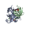
| ||||||||||
|---|---|---|---|---|---|---|---|---|---|---|---|
| 1 |
| ||||||||||
| Unit cell |
| ||||||||||
| Noncrystallographic symmetry (NCS) | NCS oper: (Code: given Matrix: (-0.99999, 0.00427, 0.00202), Vector: |
-Components
| #1: Protein | Mass: 13153.066 Da / Num. of mol.: 2 Source method: isolated from a genetically manipulated source Source: (gene. exp.) Desulfovibrio vulgaris str. 'Miyazaki F' (bacteria)Species: Desulfovibrio vulgaris / Strain: MIYAZAKI F / Cellular location: CYTOPLASM / Plasmid: PMKBT-100 / Cellular location (production host): CYTOPLASM / Production host: #2: Chemical |   Mass: 456.344 Da / Num. of mol.: 2 / Source method: obtained synthetically / Formula: C17H21N4O9P Mass: 456.344 Da / Num. of mol.: 2 / Source method: obtained synthetically / Formula: C17H21N4O9P#3: Water | ChemComp-HOH / |  Mass: 18.015 Da / Num. of mol.: 200 / Source method: isolated from a natural source / Formula: H2O Mass: 18.015 Da / Num. of mol.: 200 / Source method: isolated from a natural source / Formula: H2O |
|---|
-Experimental details
-Experiment
| Experiment | Method: X-RAY DIFFRACTION / Number of used crystals: 2 |
|---|
- Sample preparation
| Crystal | Density Matthews: 2.36 Å3/Da / Density % sol: 49.89 % | ||||||||||||||||||||||||||||||||||||||||
|---|---|---|---|---|---|---|---|---|---|---|---|---|---|---|---|---|---|---|---|---|---|---|---|---|---|---|---|---|---|---|---|---|---|---|---|---|---|---|---|---|---|
| Crystal grow | pH: 7.5 Details: 20% PEG6000, 0.1M TRIS(PH7.5), 20% GRYCEROL, 0.2M NACL | ||||||||||||||||||||||||||||||||||||||||
| Crystal grow | *PLUS pH: 8 / Method: vapor diffusion | ||||||||||||||||||||||||||||||||||||||||
| Components of the solutions | *PLUS
|
-Data collection
| Diffraction | Mean temperature: 290 K |
|---|---|
| Diffraction source | Source: SYNCHROTRON / Site: Photon Factory  / Beamline: BL-18B / Wavelength: 1 / Beamline: BL-18B / Wavelength: 1 |
| Detector | Detector: IMAGE PLATE / Date: Dec 15, 1997 / Details: BENT MIRROR |
| Radiation | Monochromator: SI(111) / Protocol: SINGLE WAVELENGTH / Monochromatic (M) / Laue (L): M / Scattering type: x-ray |
| Radiation wavelength | Wavelength: 1 Å / Relative weight: 1 |
| Reflection | Resolution: 1.2→30 Å / Num. obs: 68514 / % possible obs: 86.6 % / Observed criterion σ(I): 1 / Redundancy: 4.9 % / Rmerge(I) obs: 0.028 / Net I/σ(I): 29 |
| Reflection shell | Resolution: 1.2→1.22 Å / Rmerge(I) obs: 0.163 / Mean I/σ(I) obs: 2 / % possible all: 60.6 |
| Reflection | *PLUS Num. measured all: 337547 / Rmerge(I) obs: 0.026 |
| Reflection shell | *PLUS % possible obs: 60.6 % |
- Processing
| Software |
| |||||||||||||||||||||||||||||||||
|---|---|---|---|---|---|---|---|---|---|---|---|---|---|---|---|---|---|---|---|---|---|---|---|---|---|---|---|---|---|---|---|---|---|---|
| Refinement | Method to determine structure: MIR / Resolution: 1.3→8 Å / Cross valid method: THROUGHOUT / σ(F): 0
| |||||||||||||||||||||||||||||||||
| Refinement step | Cycle: LAST / Resolution: 1.3→8 Å
| |||||||||||||||||||||||||||||||||
| Refine LS restraints |
|
