 Movie
Movie Controller

[English] 日本語
 Yorodumi
- PDB-1wli: L122Y mutant of FMN-binding protein from Desulfovibrio vulgaris (... -
+ Open data

- Basic information
| Entry | Database: PDB / ID: 1wli | ||||||
|---|---|---|---|---|---|---|---|
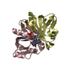
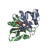
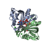
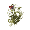
| Title | L122Y mutant of FMN-binding protein from Desulfovibrio vulgaris (Miyazaki F) | ||||||
 Components Components | FMN-binding protein | ||||||
 Keywords Keywords | ELECTRON TRANSPORT / Flavoprotein / FMN | ||||||
| Function / homology |  Function and homology information Function and homology information | ||||||
| Biological species |  Desulfovibrio vulgaris str. 'Miyazaki F' (bacteria) Desulfovibrio vulgaris str. 'Miyazaki F' (bacteria) | ||||||
| Method |  X-RAY DIFFRACTION / SYNCHROTRON / AB INITIO / Resolution: 1.6 Å X-RAY DIFFRACTION / SYNCHROTRON / AB INITIO / Resolution: 1.6 Å | ||||||
 Authors Authors | Shibata, N. / Higuchi, Y. | ||||||
 Citation Citation | Journal: J.Biochem. / Year: 2007 Title: Determination of the role of the Carboxyl-terminal leucine-122 in FMN-binding protein by mutational and structural analysis. Authors: Kitamura, M. / Terakawa, K. / Inoue, H. / Hayashida, T. / Suto, K. / Morimoto, Y. / Yasuoka, N. / Shibata, N. / Higuchi, Y. | ||||||
| History |
|
- Structure visualization
| Structure viewer | Molecule: MolmilJmol/JSmol |
|---|
- Downloads & links
-Download
| PDBx/mmCIF format | 1wli.cif.gz | 68 KB | Display | PDBx/mmCIF format |
|---|---|---|---|---|
| PDB format | pdb1wli.ent.gz | 49.2 KB | Display | PDB format |
| PDBx/mmJSON format | 1wli.json.gz | Tree view | PDBx/mmJSON format | |
| Others |  Other downloads Other downloads |
-Validation report
| Arichive directory | https://data.pdbj.org/pub/pdb/validation_reports/wl/1wliftp://data.pdbj.org/pub/pdb/validation_reports/wl/1wli | HTTPS FTP |
|---|
-Related structure data
| Related structure data |  1wlkC  3a20C  1flmS S: Starting model for refinement C: citing same article ( |
|---|---|
| Similar structure data |


-Links
 PDBj
PDBj- Assembly
| Deposited unit | 
| ||||||||
|---|---|---|---|---|---|---|---|---|---|
| 1 |
| ||||||||
| Unit cell |
|
-Components
| #1: Protein | Mass: 13203.083 Da / Num. of mol.: 2 / Mutation: L122Y Source method: isolated from a genetically manipulated source Source: (gene. exp.) Desulfovibrio vulgaris str. 'Miyazaki F' (bacteria)Species: Desulfovibrio vulgaris / Strain: Miyazaki F / Production host: #2: Chemical |   Mass: 456.344 Da / Num. of mol.: 2 / Source method: obtained synthetically / Formula: C17H21N4O9P Mass: 456.344 Da / Num. of mol.: 2 / Source method: obtained synthetically / Formula: C17H21N4O9P#3: Water | ChemComp-HOH / |  Mass: 18.015 Da / Num. of mol.: 255 / Source method: isolated from a natural source / Formula: H2O Mass: 18.015 Da / Num. of mol.: 255 / Source method: isolated from a natural source / Formula: H2O |
|---|
-Experimental details
-Experiment
| Experiment | Method: X-RAY DIFFRACTION / Number of used crystals: 1 |
|---|
- Sample preparation
| Crystal | Density Matthews: 1.89 Å3/Da / Density % sol: 35 % |
|---|---|
| Crystal grow | Temperature: 298 K / Method: vapor diffusion, sitting drop / pH: 7.5 Details: PEG6000, sodium acetate, Tris, pH 7.5, VAPOR DIFFUSION, SITTING DROP, temperature 298K |
-Data collection
| Diffraction | Mean temperature: 100 K |
|---|---|
| Diffraction source | Source: SYNCHROTRON / Site: SPring-8  / Beamline: BL44XU / Beamline: BL44XU |
| Detector | Type: OXFORD / Detector: CCD / Date: Dec 7, 2002 |
| Radiation | Protocol: SINGLE WAVELENGTH / Monochromatic (M) / Laue (L): M / Scattering type: x-ray |
| Radiation wavelength | Relative weight: 1 |
| Reflection | Highest resolution: 1.6 Å / Num. all: 29692 / Num. obs: 29692 / % possible obs: 98 % / Observed criterion σ(F): 0 / Observed criterion σ(I): -3 |
| Reflection shell | Resolution: 1.6→1.66 Å / % possible all: 84.7 |
- Processing
| Software |
| |||||||||||||||||||||||||||||||||
|---|---|---|---|---|---|---|---|---|---|---|---|---|---|---|---|---|---|---|---|---|---|---|---|---|---|---|---|---|---|---|---|---|---|---|
| Refinement | Method to determine structure: AB INITIO Starting model: 1FLM Resolution: 1.6→8 Å / Num. parameters: 8895 / Num. restraintsaints: 8133 / Cross valid method: THROUGHOUT / σ(F): 0 / Stereochemistry target values: ENGH & HUBER
| |||||||||||||||||||||||||||||||||
| Refine analyze | Num. disordered residues: 5 / Occupancy sum hydrogen: 0 / Occupancy sum non hydrogen: 2177 | |||||||||||||||||||||||||||||||||
| Refinement step | Cycle: LAST / Resolution: 1.6→8 Å
| |||||||||||||||||||||||||||||||||
| Refine LS restraints |
| |||||||||||||||||||||||||||||||||
| LS refinement shell | Resolution: 1.6→1.66 Å / Rfactor Rwork: 0.157 |
