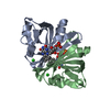
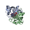
STRUCTURAL GENOMICS / UNKNOWN FUNCTION / conserved hypothetical protein YDCE / New York SGX Research Center for Structural Genomics / PSI / Protein Structure Initiative / NYSGXRC
Function / homology
Function and homology information
rRNA catabolic process / Hydrolases; Acting on ester bonds; Endoribonucleases producing 3'-phosphomonoesters / mRNA catabolic process / RNA endonuclease activity / hydrolase activity / DNA binding / RNA binding Similarity search - Function
SH3 type barrels. - #110 / mRNA interferase PemK-like / PemK-like, MazF-like toxin of type II toxin-antitoxin system / Plasmid maintenance toxin/Cell growth inhibitor / SH3 type barrels. / Roll / Mainly Beta Similarity search - Domain/homology
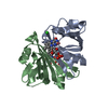
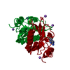
The second molecule of the putative biological dimer is generated by the following transformation: BIOMT1 0.5 -0.866 0.0 28.3175 BIOMT2 -0.866 -0.5 0.0 49.045 BIOMT3 0.0 0.0 -1.0 23.0431
-
Components
#1: Protein
conservedhypotheticalproteinYDCE
Mass: 13062.981 Da / Num. of mol.: 1 Source method: isolated from a genetically manipulated source Source: (gene. exp.) Bacillus subtilis (bacteria) / Gene: ydcE / Plasmid: pSMT3 / Species (production host): Escherichia coli / Production host: Escherichia coli BL21 (bacteria) / Strain (production host): BL21 / References: UniProt: P96622
In the structure databanks used in Yorodumi, some data are registered as the other names, "COVID-19 virus" and "2019-nCoV". Here are the details of the virus and the list of structure data.
Jan 31, 2019. EMDB accession codes are about to change! (news from PDBe EMDB page)
EMDB accession codes are about to change! (news from PDBe EMDB page)
The allocation of 4 digits for EMDB accession codes will soon come to an end. Whilst these codes will remain in use, new EMDB accession codes will include an additional digit and will expand incrementally as the available range of codes is exhausted. The current 4-digit format prefixed with “EMD-” (i.e. EMD-XXXX) will advance to a 5-digit format (i.e. EMD-XXXXX), and so on. It is currently estimated that the 4-digit codes will be depleted around Spring 2019, at which point the 5-digit format will come into force.
The EM Navigator/Yorodumi systems omit the EMD- prefix.
Related info.:Q: What is EMD? / ID/Accession-code notation in Yorodumi/EM Navigator
Yorodumi is a browser for structure data from EMDB, PDB, SASBDB, etc.
This page is also the successor to EM Navigator detail page, and also detail information page/front-end page for Omokage search.
The word "yorodu" (or yorozu) is an old Japanese word meaning "ten thousand". "mi" (miru) is to see.
Related info.:EMDB / PDB / SASBDB / Comparison of 3 databanks / Yorodumi Search / Aug 31, 2016. New EM Navigator & Yorodumi / Yorodumi Papers / Jmol/JSmol / Function and homology information / Changes in new EM Navigator and Yorodumi
 Movie
Movie Controller


 Open data
 Basic information
 Components
 Keywords
 Function and homology information
 X-RAY DIFFRACTION /
  Authors
 Citation
 Structure visualization
 Downloads & links
 Other downloads
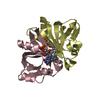

 PDBj
PDBj

 Assembly
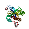

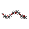
 Mass: 252.305 Da / Num. of mol.: 2 / Source method: obtained synthetically / Formula: C11H24O6
 Mass: 60.052 Da / Num. of mol.: 2 / Source method: obtained synthetically / Formula: C2H4O2
 Mass: 18.015 Da / Num. of mol.: 100 / Source method: isolated from a natural source / Formula: H2O
 Sample preparation
 / Beamline: X9A / Wavelength: 0.9790, 0.97938, 0.97163
 Processing