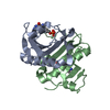
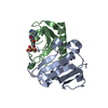
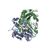
 Movie
Movie Controller


+ Open data

- Basic information
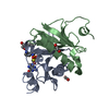
| Entry | Database: PDB / ID: 3n1s | ||||||
|---|---|---|---|---|---|---|---|
| Title | Crystal structure of wild type ecHint GMP complex | ||||||
 Components Components | HIT-like protein hinT | ||||||
 Keywords Keywords | HYDROLASE / histidine triad nucleotide binding protein / Hint / GMP | ||||||
| Function / homology |  Function and homology information Function and homology informationD-alanine catabolic process / Hydrolases; Acting on phosphorus-nitrogen bonds / adenosine 5'-monophosphoramidase activity / nucleotide binding / protein homodimerization activity / cytoplasm / cytosol Similarity search - Function | ||||||
| Biological species |  | ||||||
| Method |  X-RAY DIFFRACTION / SYNCHROTRON / MOLECULAR REPLACEMENT / Resolution: 1.45 Å X-RAY DIFFRACTION / SYNCHROTRON / MOLECULAR REPLACEMENT / Resolution: 1.45 Å | ||||||
 Authors Authors | Cody, V. | ||||||
 Citation Citation | Journal: J.Mol.Biol. / Year: 2010 Title: Probing the Impact of the echinT C-Terminal Domain on Structure and Catalysis. Authors: Bardaweel, S. / Pace, J. / Chou, T.F. / Cody, V. / Wagner, C.R. | ||||||
| History |
|
- Structure visualization
| Structure viewer | Molecule: MolmilJmol/JSmol |
|---|
- Downloads & links
-Download
| PDBx/mmCIF format | 3n1s.cif.gz | 230.4 KB | Display | PDBx/mmCIF format |
|---|---|---|---|---|
| PDB format | pdb3n1s.ent.gz | 185.6 KB | Display | PDB format |
| PDBx/mmJSON format | 3n1s.json.gz | Tree view | PDBx/mmJSON format | |
| Others |  Other downloads Other downloads |
-Validation report
| Arichive directory | https://data.pdbj.org/pub/pdb/validation_reports/n1/3n1sftp://data.pdbj.org/pub/pdb/validation_reports/n1/3n1s | HTTPS FTP |
|---|
-Related structure data
| Related structure data |  3n1tC  1av5S S: Starting model for refinement C: citing same article ( |
|---|---|
| Similar structure data |


-Links
 PDBj
PDBj


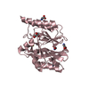
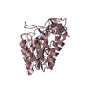
- Assembly
| Deposited unit | 
| ||||||||
|---|---|---|---|---|---|---|---|---|---|
| 1 | 
| ||||||||
| 2 | 
| ||||||||
| 3 | 
| ||||||||
| 4 | 
| ||||||||
| Unit cell |
|
-Components
| #1: Protein | Mass: 13258.262 Da / Num. of mol.: 8 Source method: isolated from a genetically manipulated source Source: (gene. exp.) #2: Chemical | ChemComp-5GP /   Mass: 363.221 Da / Num. of mol.: 8 / Source method: obtained synthetically / Formula: C10H14N5O8P Mass: 363.221 Da / Num. of mol.: 8 / Source method: obtained synthetically / Formula: C10H14N5O8P#3: Chemical | ChemComp-EDO /   Mass: 62.068 Da / Num. of mol.: 25 / Source method: obtained synthetically / Formula: C2H6O2 Mass: 62.068 Da / Num. of mol.: 25 / Source method: obtained synthetically / Formula: C2H6O2#4: Water | ChemComp-HOH / |  Mass: 18.015 Da / Num. of mol.: 1068 / Source method: isolated from a natural source / Formula: H2O Mass: 18.015 Da / Num. of mol.: 1068 / Source method: isolated from a natural source / Formula: H2O |
|---|
-Experimental details
-Experiment
| Experiment | Method: X-RAY DIFFRACTION / Number of used crystals: 1 |
|---|
- Sample preparation
| Crystal | Density Matthews: 2.16 Å3/Da / Density % sol: 43.03 % |
|---|---|
| Crystal grow | Temperature: 298 K / Method: vapor diffusion, hanging drop / pH: 7 Details: 40% PEG 20000, 0.1M Magnesium Acetate, 0.1M Sodium Acetate, 20 mM Tris, pH 7.0, 1mM EDTA, 10% glycerol, VAPOR DIFFUSION, HANGING DROP, temperature 298K |
-Data collection
| Diffraction | Mean temperature: 113 K |
|---|---|
| Diffraction source | Source: SYNCHROTRON / Site: SSRL  / Beamline: BL9-1 / Wavelength: 0.975 Å / Beamline: BL9-1 / Wavelength: 0.975 Å |
| Detector | Type: MARMOSAIC 325 mm CCD / Detector: CCD / Date: Jun 6, 2006 / Details: mirrors |
| Radiation | Monochromator: graphite / Protocol: SINGLE WAVELENGTH / Monochromatic (M) / Laue (L): M / Scattering type: x-ray |
| Radiation wavelength | Wavelength: 0.975 Å / Relative weight: 1 |
| Reflection | Resolution: 1.35→38 Å / Num. all: 222096 / Num. obs: 149986 / % possible obs: 95.2 % / Observed criterion σ(F): 2 / Observed criterion σ(I): 2 / Redundancy: 3.4 % / Biso Wilson estimate: 20.4 Å2 / Rmerge(I) obs: 0.165 / Rsym value: 0.115 / Net I/σ(I): 5.3 |
| Reflection shell | Resolution: 1.35→1.45 Å / Redundancy: 2.5 % / Rmerge(I) obs: 0.215 / Mean I/σ(I) obs: 3.2 / Num. unique all: 17208 / Rsym value: 0.4 / % possible all: 77.7 |
- Processing
| Software |
| ||||||||||||||||||||||||||||||||||||||||||||||||||||||||||||||||||||||||||||||||||||||||||
|---|---|---|---|---|---|---|---|---|---|---|---|---|---|---|---|---|---|---|---|---|---|---|---|---|---|---|---|---|---|---|---|---|---|---|---|---|---|---|---|---|---|---|---|---|---|---|---|---|---|---|---|---|---|---|---|---|---|---|---|---|---|---|---|---|---|---|---|---|---|---|---|---|---|---|---|---|---|---|---|---|---|---|---|---|---|---|---|---|---|---|---|
| Refinement | Method to determine structure: MOLECULAR REPLACEMENT Starting model: PDB ENTRY 1AV5 Resolution: 1.45→37.96 Å / Cor.coef. Fo:Fc: 0.97 / Cor.coef. Fo:Fc free: 0.96 / SU B: 1.299 / SU ML: 0.051 / Cross valid method: THROUGHOUT / ESU R: 0.074 / ESU R Free: 0.076 / Stereochemistry target values: MAXIMUM LIKELIHOOD / Details: HYDROGENS HAVE BEEN ADDED IN THE RIDING POSITIONS
| ||||||||||||||||||||||||||||||||||||||||||||||||||||||||||||||||||||||||||||||||||||||||||
| Solvent computation | Ion probe radii: 0.8 Å / Shrinkage radii: 0.8 Å / VDW probe radii: 1.2 Å / Solvent model: MASK | ||||||||||||||||||||||||||||||||||||||||||||||||||||||||||||||||||||||||||||||||||||||||||
| Displacement parameters | Biso mean: 20.504 Å2
| ||||||||||||||||||||||||||||||||||||||||||||||||||||||||||||||||||||||||||||||||||||||||||
| Refine analyze | Luzzati coordinate error obs: 0.163 Å | ||||||||||||||||||||||||||||||||||||||||||||||||||||||||||||||||||||||||||||||||||||||||||
| Refinement step | Cycle: LAST / Resolution: 1.45→37.96 Å
| ||||||||||||||||||||||||||||||||||||||||||||||||||||||||||||||||||||||||||||||||||||||||||
| Refine LS restraints |
| ||||||||||||||||||||||||||||||||||||||||||||||||||||||||||||||||||||||||||||||||||||||||||
| LS refinement shell | Resolution: 1.45→1.488 Å / Total num. of bins used: 20
|
