- PDB-3dmb: Crystal structure of a putative general stress family protein (xc... -
+
Open data
ID or keywords:
Loading...
-
Basic information
Entry
Database: PDB / ID: 3dmb
Title
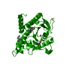
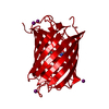
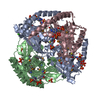
Crystal structure of a putative general stress family protein (xcc2264) from xanthomonas campestris pv. campestris at 2.30 A resolution
Components
Putative General Stress Protein 26 with a PNP-Oxidase like Fold
Keywords
OXIDOREDUCTASE / Pnp-oxidase like fold / structural genomics / Joint Center for Structural Genomics / JCSG / Protein Structure Initiative / PSI-2
Function / homology
General stress protein, FMN-binding split barrel domain / Pyridoxamine 5'-phosphate oxidase like / : / Electron Transport, Fmn-binding Protein; Chain A / Pnp Oxidase; Chain A / FMN-binding split barrel / Roll / Mainly Beta / General stress protein
Function and homology information
Biological species
Xanthomonas campestris pv. campestris (bacteria)
Method
X-RAY DIFFRACTION / SYNCHROTRON / MAD / Resolution: 2.3 Å
A: Putative General Stress Protein 26 with a PNP-Oxidase like Fold B: Putative General Stress Protein 26 with a PNP-Oxidase like Fold C: Putative General Stress Protein 26 with a PNP-Oxidase like Fold
A: Putative General Stress Protein 26 with a PNP-Oxidase like Fold B: Putative General Stress Protein 26 with a PNP-Oxidase like Fold C: Putative General Stress Protein 26 with a PNP-Oxidase like Fold
A: Putative General Stress Protein 26 with a PNP-Oxidase like Fold B: Putative General Stress Protein 26 with a PNP-Oxidase like Fold C: Putative General Stress Protein 26 with a PNP-Oxidase like Fold
Mass: 18.015 Da / Num. of mol.: 64 / Source method: isolated from a natural source / Formula: H2O
Has protein modification
Y
Sequence details
THE CONSTRUCT CONTAINS AMINO ACIDS 1-146 OF THE FULL LENGTH PROTEIN OF 162 RESIDUES. THE CONSTRUCT ...THE CONSTRUCT CONTAINS AMINO ACIDS 1-146 OF THE FULL LENGTH PROTEIN OF 162 RESIDUES. THE CONSTRUCT WAS EXPRESSED WITH A PURIFICATION TAG MGSDKIHHHHHHENLYFQG. THE TAG WAS REMOVED WITH TEV PROTEASE LEAVING ONLY A GLYCINE (0) FOLLOWED BY THE TARGET SEQUENCE.
-
Experimental details
-
Experiment
Experiment
Method: X-RAY DIFFRACTION / Number of used crystals: 1
-
Sample preparation
Crystal
Density Matthews: 2.26 Å3/Da / Density % sol: 45.56 %
Crystal grow
Temperature: 277 K / Method: vapor diffusion, sitting drop / pH: 4.5 Details: 1.2000M K2HPO4, 0.8000M Na2HPO4, 0.1M Acetate pH 4.5, NANODROP, VAPOR DIFFUSION, SITTING DROP, temperature 277K
Type: MARMOSAIC 325 mm CCD / Detector: CCD / Date: May 16, 2008 / Details: Flat mirror (vertical focusing)
Radiation
Monochromator: Single crystal Si(111) bent monochromator (horizontal focusing) Protocol: MAD / Monochromatic (M) / Laue (L): M / Scattering type: x-ray
Radiation wavelength
ID
Wavelength (Å)
Relative weight
1
0.97862
1
2
0.91837
1
3
0.97939
1
Reflection
Resolution: 2.3→28.665 Å / Num. obs: 19745 / % possible obs: 99.4 % / Observed criterion σ(I): -3 / Biso Wilson estimate: 52.88 Å2 / Rmerge(I) obs: 0.048 / Net I/σ(I): 17.56
Reflection shell
Diffraction-ID: 1
Resolution (Å)
Highest resolution (Å)
Rmerge(I) obs
Mean I/σ(I) obs
Num. measured obs
Num. unique obs
% possible all
2.3-2.38
0.642
2.1
13091
3545
96
2.38-2.48
0.496
2.8
14906
3915
99.7
2.48-2.59
0.377
3.8
14154
3702
99.9
2.59-2.73
0.29
4.8
14776
3858
99.8
2.73-2.9
0.177
7.7
14203
3718
99.8
2.9-3.12
0.102
12.5
14413
3748
99.7
3.12-3.43
0.06
19.7
14357
3738
99.9
3.43-3.93
0.035
30.1
14711
3825
99.9
3.93-4.93
0.023
43.2
14440
3739
99.9
4.93
0.021
48.1
14717
3827
99.3
-
Phasing
Phasing
Method: MAD
-
Processing
Software
Name
Version
Classification
NB
REFMAC
5.2.0019
refinement
PHENIX
refinement
SHELX
phasing
MolProbity
3beta29
modelbuilding
XSCALE
datascaling
PDB_EXTRACT
3.004
dataextraction
XDS
datareduction
SHELXD
phasing
autoSHARP
phasing
Refinement
Method to determine structure: MAD / Resolution: 2.3→28.665 Å / Cor.coef. Fo:Fc: 0.951 / Cor.coef. Fo:Fc free: 0.924 / SU B: 18.407 / SU ML: 0.216 / TLS residual ADP flag: LIKELY RESIDUAL / Cross valid method: THROUGHOUT / σ(F): 0 / ESU R: 0.381 / ESU R Free: 0.26 Stereochemistry target values: MAXIMUM LIKELIHOOD WITH PHASES Details: 1. HYDROGENS HAVE BEEN ADDED IN THE RIDING POSITIONS. 2. ATOM RECORDS CONTAIN RESIDUAL B FACTORS ONLY. 3. A MET-INHIBITION PROTOCOL WAS USED FOR SELENOMETHIONINE INCORPORATION DURING PROTEIN ...Details: 1. HYDROGENS HAVE BEEN ADDED IN THE RIDING POSITIONS. 2. ATOM RECORDS CONTAIN RESIDUAL B FACTORS ONLY. 3. A MET-INHIBITION PROTOCOL WAS USED FOR SELENOMETHIONINE INCORPORATION DURING PROTEIN EXPRESSION. THE OCCUPANCY OF THE SE ATOMS IN THE MSE RESIDUES WAS REDUCED TO 0.75 FOR THE REDUCED SCATTERING POWER DUE TO PARTIAL S-MET INCORPORATION.
Rfactor
Num. reflection
% reflection
Selection details
Rfree
0.264
1006
5.1 %
RANDOM
Rwork
0.21
-
-
-
obs
0.213
19719
99.7 %
-
Solvent computation
Ion probe radii: 0.8 Å / Shrinkage radii: 0.8 Å / VDW probe radii: 1.2 Å / Solvent model: BABINET MODEL WITH MASK
Displacement parameters
Biso mean: 56.5 Å2
Baniso -1
Baniso -2
Baniso -3
1-
1.08 Å2
0.54 Å2
0 Å2
2-
-
1.08 Å2
0 Å2
3-
-
-
-1.61 Å2
Refinement step
Cycle: LAST / Resolution: 2.3→28.665 Å
Protein
Nucleic acid
Ligand
Solvent
Total
Num. atoms
3219
0
0
64
3283
Refine LS restraints
Refine-ID
Type
Dev ideal
Dev ideal target
Number
X-RAY DIFFRACTION
r_bond_refined_d
0.014
0.022
3316
X-RAY DIFFRACTION
r_bond_other_d
0.003
0.02
2241
X-RAY DIFFRACTION
r_angle_refined_deg
1.671
1.957
4496
X-RAY DIFFRACTION
r_angle_other_deg
1.281
3
5460
X-RAY DIFFRACTION
r_dihedral_angle_1_deg
3.749
5
429
X-RAY DIFFRACTION
r_dihedral_angle_2_deg
34.754
24.161
137
X-RAY DIFFRACTION
r_dihedral_angle_3_deg
13.173
15
544
X-RAY DIFFRACTION
r_dihedral_angle_4_deg
10.207
15
18
X-RAY DIFFRACTION
r_chiral_restr
0.096
0.2
488
X-RAY DIFFRACTION
r_gen_planes_refined
0.005
0.02
3733
X-RAY DIFFRACTION
r_gen_planes_other
0.002
0.02
670
X-RAY DIFFRACTION
r_nbd_refined
0.162
0.2
528
X-RAY DIFFRACTION
r_nbd_other
0.138
0.2
1991
X-RAY DIFFRACTION
r_nbtor_refined
0.15
0.2
1530
X-RAY DIFFRACTION
r_nbtor_other
0.07
0.2
1770
X-RAY DIFFRACTION
r_xyhbond_nbd_refined
0.11
0.2
88
X-RAY DIFFRACTION
r_symmetry_vdw_refined
0.161
0.2
32
X-RAY DIFFRACTION
r_symmetry_vdw_other
0.124
0.2
107
X-RAY DIFFRACTION
r_symmetry_hbond_refined
0.077
0.2
14
X-RAY DIFFRACTION
r_mcbond_it
1.125
2
2402
X-RAY DIFFRACTION
r_mcbond_other
0.235
2
883
X-RAY DIFFRACTION
r_mcangle_it
1.763
4
3328
X-RAY DIFFRACTION
r_scbond_it
3.073
6
1391
X-RAY DIFFRACTION
r_scangle_it
4.44
8
1164
Refine LS restraints NCS
Ens-ID: 1 / Refine-ID: X-RAY DIFFRACTION
Dom-ID
Auth asym-ID
Number
Type
Rms dev position (Å)
Weight position
1
A
694
MEDIUMPOSITIONAL
0.35
0.5
2
B
694
MEDIUMPOSITIONAL
0.33
0.5
3
C
694
MEDIUMPOSITIONAL
0.28
0.5
1
A
807
LOOSEPOSITIONAL
0.83
5
2
B
807
LOOSEPOSITIONAL
0.66
5
3
C
807
LOOSEPOSITIONAL
0.67
5
1
A
694
MEDIUMTHERMAL
0.79
2
2
B
694
MEDIUMTHERMAL
0.74
2
3
C
694
MEDIUMTHERMAL
0.71
2
1
A
807
LOOSETHERMAL
2.18
10
2
B
807
LOOSETHERMAL
1.82
10
3
C
807
LOOSETHERMAL
1.84
10
LS refinement shell
Resolution: 2.299→2.359 Å / Total num. of bins used: 20
Rfactor
Num. reflection
% reflection
Rfree
0.305
74
-
Rwork
0.281
1353
-
all
-
1427
-
obs
-
-
98.55 %
Refinement TLS params.
Method: refined / Refine-ID: X-RAY DIFFRACTION
ID
L11 (°2)
L12 (°2)
L13 (°2)
L22 (°2)
L23 (°2)
L33 (°2)
S11 (Å °)
S12 (Å °)
S13 (Å °)
S21 (Å °)
S22 (Å °)
S23 (Å °)
S31 (Å °)
S32 (Å °)
S33 (Å °)
T11 (Å2)
T12 (Å2)
T13 (Å2)
T22 (Å2)
T23 (Å2)
T33 (Å2)
Origin x (Å)
Origin y (Å)
Origin z (Å)
1
8.2469
1.4722
1.0181
0.9783
0.3035
3.4273
0.0224
0.2345
-0.208
-0.0659
0.0091
0.0363
0.2098
-0.105
-0.0314
-0.1911
0.0192
-0.0078
-0.2845
-0.0069
-0.2973
6.838
22.919
15.748
2
3.1748
0.0879
1.8921
0.9104
-0.445
4.7071
-0.0338
0.2244
-0.0845
-0.0736
-0.0107
-0.2336
0.1496
0.8817
0.0445
-0.1705
0.0576
0.058
-0.0756
0.0295
-0.1589
27.241
21.269
16.909
3
0.3114
0.8513
-0.089
3.0589
2.2963
8.8336
0.3686
-0.0292
0.1318
0.3207
-0.3648
0.0896
-1.8754
-0.0964
-0.0037
0.4092
-0.2007
0.0293
-0.2317
-0.0078
-0.0864
23.357
52.43
9.367
Refinement TLS group
ID
Refine-ID
Refine TLS-ID
Auth asym-ID
Label asym-ID
Auth seq-ID
Label seq-ID
1
X-RAY DIFFRACTION
1
A
A
0 - 145
1 - 146
2
X-RAY DIFFRACTION
2
B
B
1 - 145
2 - 146
3
X-RAY DIFFRACTION
3
C
C
4 - 24
5 - 25
4
X-RAY DIFFRACTION
3
C
C
33 - 145
34 - 146
+
About Yorodumi
-
News
-
Feb 9, 2022. New format data for meta-information of EMDB entries
New format data for meta-information of EMDB entries
Version 3 of the EMDB header file is now the official format.
The previous official version 1.9 will be removed from the archive.
In the structure databanks used in Yorodumi, some data are registered as the other names, "COVID-19 virus" and "2019-nCoV". Here are the details of the virus and the list of structure data.
Jan 31, 2019. EMDB accession codes are about to change! (news from PDBe EMDB page)
EMDB accession codes are about to change! (news from PDBe EMDB page)
The allocation of 4 digits for EMDB accession codes will soon come to an end. Whilst these codes will remain in use, new EMDB accession codes will include an additional digit and will expand incrementally as the available range of codes is exhausted. The current 4-digit format prefixed with “EMD-” (i.e. EMD-XXXX) will advance to a 5-digit format (i.e. EMD-XXXXX), and so on. It is currently estimated that the 4-digit codes will be depleted around Spring 2019, at which point the 5-digit format will come into force.
The EM Navigator/Yorodumi systems omit the EMD- prefix.
Related info.:Q: What is EMD? / ID/Accession-code notation in Yorodumi/EM Navigator
Yorodumi is a browser for structure data from EMDB, PDB, SASBDB, etc.
This page is also the successor to EM Navigator detail page, and also detail information page/front-end page for Omokage search.
The word "yorodu" (or yorozu) is an old Japanese word meaning "ten thousand". "mi" (miru) is to see.
Related info.:EMDB / PDB / SASBDB / Comparison of 3 databanks / Yorodumi Search / Aug 31, 2016. New EM Navigator & Yorodumi / Yorodumi Papers / Jmol/JSmol / Function and homology information / Changes in new EM Navigator and Yorodumi
 Movie
Movie Controller

 Yorodumi
 Open data
 Basic information
 Components
 Keywords
 Function and homology information
 Xanthomonas campestris pv. campestris (bacteria)
 X-RAY DIFFRACTION /
  Authors
 Citation
 Structure visualization
 Downloads & links
 Other downloads
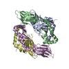
 PDBj
PDBj Assembly

 Mass: 18.015 Da / Num. of mol.: 64 / Source method: isolated from a natural source / Formula: H2O
 Sample preparation
 / Beamline: BL11-1 / Wavelength: 0.97862,0.91837,0.97939
 Processing