 Movie
Movie Controller

[English] 日本語
 Yorodumi
- PDB-5xyo: Structure of 6-aminohexanoate-oligomer hydrolase from Arthrobacte... -
+ Open data

- Basic information
| Entry | Database: PDB / ID: 5xyo | ||||||
|---|---|---|---|---|---|---|---|
| Title | Structure of 6-aminohexanoate-oligomer hydrolase from Arthrobacter sp. KI72., D122G mutant | ||||||
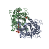
 Components Components | Endo-type 6-aminohexanoate oligomer hydrolase | ||||||
 Keywords Keywords | HYDROLASE / Nylon oligomer | ||||||
| Function / homology |  Function and homology information Function and homology information6-aminohexanoate-oligomer endohydrolase / nylon catabolic process / aminopeptidase activity Similarity search - Function | ||||||
| Biological species |  Flavobacterium sp. KI723T1 (bacteria) Flavobacterium sp. KI723T1 (bacteria) | ||||||
| Method |  X-RAY DIFFRACTION / MOLECULAR REPLACEMENT / Resolution: 2 Å X-RAY DIFFRACTION / MOLECULAR REPLACEMENT / Resolution: 2 Å | ||||||
 Authors Authors | Negoro, S. / Shibata, N. / Nagai, K. / Higuchi, Y. | ||||||
 Citation Citation | Journal: Sci Rep / Year: 2018 Title: Structural basis of the correct subunit assembly, aggregation, and intracellular degradation of nylon hydrolase Authors: Negoro, S. / Shibata, N. / Lee, Y.H. / Takehara, I. / Kinugasa, R. / Nagai, K. / Tanaka, Y. / Kato, D.I. / Takeo, M. / Goto, Y. / Higuchi, Y. | ||||||
| History |
|
- Structure visualization
| Structure viewer | Molecule: MolmilJmol/JSmol |
|---|
- Downloads & links
-Download
| PDBx/mmCIF format | 5xyo.cif.gz | 254.2 KB | Display | PDBx/mmCIF format |
|---|---|---|---|---|
| PDB format | pdb5xyo.ent.gz | 204 KB | Display | PDB format |
| PDBx/mmJSON format | 5xyo.json.gz | Tree view | PDBx/mmJSON format | |
| Others |  Other downloads Other downloads |
-Validation report
| Arichive directory | https://data.pdbj.org/pub/pdb/validation_reports/xy/5xyoftp://data.pdbj.org/pub/pdb/validation_reports/xy/5xyo | HTTPS FTP |
|---|
-Related structure data
| Related structure data | 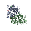 5xygSC  5xypC 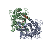 5xyqC 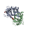 5xysC 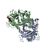 5xytC 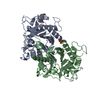 5y0lC 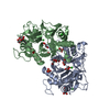 5y0mC S: Starting model for refinement C: citing same article ( |
|---|---|
| Similar structure data |


-Links
 PDBj
PDBj- Assembly
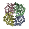
| Deposited unit | 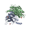
| |||||||||||||||||||||||||||||||||||||||
|---|---|---|---|---|---|---|---|---|---|---|---|---|---|---|---|---|---|---|---|---|---|---|---|---|---|---|---|---|---|---|---|---|---|---|---|---|---|---|---|---|
| 1 | 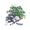
| |||||||||||||||||||||||||||||||||||||||
| Unit cell |
| |||||||||||||||||||||||||||||||||||||||
| Noncrystallographic symmetry (NCS) | NCS domain:
NCS domain segments: End auth comp-ID: LYS / End label comp-ID: LYS / Refine code: 5
NCS ensembles :
|
-Components
| #1: Protein | Mass: 36881.199 Da / Num. of mol.: 2 / Mutation: D122G Source method: isolated from a genetically manipulated source Source: (gene. exp.) Flavobacterium sp. KI723T1 (bacteria) / Gene: nylC / Production host: #2: Chemical |   Mass: 96.063 Da / Num. of mol.: 2 / Source method: obtained synthetically / Formula: SO4 Mass: 96.063 Da / Num. of mol.: 2 / Source method: obtained synthetically / Formula: SO4#3: Chemical |   Mass: 35.453 Da / Num. of mol.: 2 / Source method: obtained synthetically / Formula: Cl Mass: 35.453 Da / Num. of mol.: 2 / Source method: obtained synthetically / Formula: Cl#4: Chemical | ChemComp-GOL / |   Mass: 92.094 Da / Num. of mol.: 1 / Source method: obtained synthetically / Formula: C3H8O3 Mass: 92.094 Da / Num. of mol.: 1 / Source method: obtained synthetically / Formula: C3H8O3#5: Water | ChemComp-HOH / |  Mass: 18.015 Da / Num. of mol.: 317 / Source method: isolated from a natural source / Formula: H2O Mass: 18.015 Da / Num. of mol.: 317 / Source method: isolated from a natural source / Formula: H2OSequence details | Authors state that the long gap between the residues is due to the post-translational auto-cleavage ...Authors state that the long gap between the residues is due to the post-translational auto-cleavage of the nascent polypeptide between Asn266 and Thr267. | |
|---|
-Experimental details
-Experiment
| Experiment | Method: X-RAY DIFFRACTION / Number of used crystals: 1 |
|---|
- Sample preparation
| Crystal | Density Matthews: 2.21 Å3/Da / Density % sol: 44.46 % |
|---|---|
| Crystal grow | Temperature: 293 K / Method: vapor diffusion, sitting drop Details: 1.2-2.2 M ammonium sulphate, 0.2 M NaCl, 0.1 M HEPES buffer pH 7.5-8.5 |
-Data collection
| Diffraction | Mean temperature: 100 K |
|---|---|
| Diffraction source | Source: ROTATING ANODE / Type: RIGAKU MICROMAX-007 / Wavelength: 1.5418 Å |
| Detector | Type: RIGAKU RAXIS VII / Detector: IMAGE PLATE / Date: Nov 11, 2008 |
| Radiation | Protocol: SINGLE WAVELENGTH / Monochromatic (M) / Laue (L): M / Scattering type: x-ray |
| Radiation wavelength | Wavelength: 1.5418 Å / Relative weight: 1 |
| Reflection | Resolution: 2→100 Å / Num. obs: 43634 / % possible obs: 97.8 % / Redundancy: 2.79 % / Rmerge(I) obs: 0.052 / Net I/σ(I): 12.9 |
| Reflection shell | Resolution: 2→2.07 Å / Redundancy: 2.05 % / Rmerge(I) obs: 0.19 / Mean I/σ(I) obs: 4.2 / % possible all: 84 |
- Processing
| Software |
| ||||||||||||||||||||||||||||||||||||||||||||||||||||||||||||||||||||||||||||||||||||||||||||||||||||||||||||||||||||||||||||||||||||||||||||||||||||||||||||||||||||||||||||||||||||||
|---|---|---|---|---|---|---|---|---|---|---|---|---|---|---|---|---|---|---|---|---|---|---|---|---|---|---|---|---|---|---|---|---|---|---|---|---|---|---|---|---|---|---|---|---|---|---|---|---|---|---|---|---|---|---|---|---|---|---|---|---|---|---|---|---|---|---|---|---|---|---|---|---|---|---|---|---|---|---|---|---|---|---|---|---|---|---|---|---|---|---|---|---|---|---|---|---|---|---|---|---|---|---|---|---|---|---|---|---|---|---|---|---|---|---|---|---|---|---|---|---|---|---|---|---|---|---|---|---|---|---|---|---|---|---|---|---|---|---|---|---|---|---|---|---|---|---|---|---|---|---|---|---|---|---|---|---|---|---|---|---|---|---|---|---|---|---|---|---|---|---|---|---|---|---|---|---|---|---|---|---|---|---|---|
| Refinement | Method to determine structure: MOLECULAR REPLACEMENT Starting model: 5XYG Resolution: 2→72.23 Å / Cor.coef. Fo:Fc: 0.961 / Cor.coef. Fo:Fc free: 0.934 / SU B: 10.057 / SU ML: 0.122 / Cross valid method: THROUGHOUT / ESU R: 0.18 / ESU R Free: 0.165
| ||||||||||||||||||||||||||||||||||||||||||||||||||||||||||||||||||||||||||||||||||||||||||||||||||||||||||||||||||||||||||||||||||||||||||||||||||||||||||||||||||||||||||||||||||||||
| Solvent computation | Ion probe radii: 0.8 Å / Shrinkage radii: 0.8 Å / VDW probe radii: 1.4 Å | ||||||||||||||||||||||||||||||||||||||||||||||||||||||||||||||||||||||||||||||||||||||||||||||||||||||||||||||||||||||||||||||||||||||||||||||||||||||||||||||||||||||||||||||||||||||
| Displacement parameters | Biso mean: 27.022 Å2
| ||||||||||||||||||||||||||||||||||||||||||||||||||||||||||||||||||||||||||||||||||||||||||||||||||||||||||||||||||||||||||||||||||||||||||||||||||||||||||||||||||||||||||||||||||||||
| Refinement step | Cycle: 1 / Resolution: 2→72.23 Å
| ||||||||||||||||||||||||||||||||||||||||||||||||||||||||||||||||||||||||||||||||||||||||||||||||||||||||||||||||||||||||||||||||||||||||||||||||||||||||||||||||||||||||||||||||||||||
| Refine LS restraints |
|