 Movie
Movie Controller


+ Open data

- Basic information
| Entry | Database: PDB / ID: 7rpy | ||||||
|---|---|---|---|---|---|---|---|
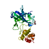
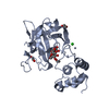
| Title | X25-2 domain of Sca5 from Ruminococcus bromii | ||||||
 Components Components | Cohesin-containing protein | ||||||
 Keywords Keywords | SUGAR BINDING PROTEIN / Starch-binding protein domain in the Ruminococcus bromii amylosome protein Sca5 | ||||||
| Function / homology | Immunoglobulins / Immunoglobulin-like / Sandwich / Mainly Beta / alpha-maltotriose / ACETATE ION Function and homology information Function and homology information | ||||||
| Biological species |  Ruminococcus bromii L2-63 (bacteria) Ruminococcus bromii L2-63 (bacteria) | ||||||
| Method |  X-RAY DIFFRACTION / SYNCHROTRON / SAD / Resolution: 1.67 Å X-RAY DIFFRACTION / SYNCHROTRON / SAD / Resolution: 1.67 Å | ||||||
 Authors Authors | Cerqueira, F. / Koropatkin, N. | ||||||
| Funding support | 1items
| ||||||
 Citation Citation | Journal: J.Biol.Chem. / Year: 2022 Title: Sas20 is a highly flexible starch-binding protein in the Ruminococcus bromii cell-surface amylosome. Authors: Cerqueira, F.M. / Photenhauer, A.L. / Doden, H.L. / Brown, A.N. / Abdel-Hamid, A.M. / Morais, S. / Bayer, E.A. / Wawrzak, Z. / Cann, I. / Ridlon, J.M. / Hopkins, J.B. / Koropatkin, N.M. | ||||||
| History |
|
- Structure visualization
| Structure viewer | Molecule: MolmilJmol/JSmol |
|---|
- Downloads & links
-Download
| PDBx/mmCIF format | 7rpy.cif.gz | 119.3 KB | Display | PDBx/mmCIF format |
|---|---|---|---|---|
| PDB format | pdb7rpy.ent.gz | 90.9 KB | Display | PDB format |
| PDBx/mmJSON format | 7rpy.json.gz | Tree view | PDBx/mmJSON format | |
| Others |  Other downloads Other downloads |
-Validation report
| Arichive directory | https://data.pdbj.org/pub/pdb/validation_reports/rp/7rpyftp://data.pdbj.org/pub/pdb/validation_reports/rp/7rpy | HTTPS FTP |
|---|
-Related structure data
-Links
 PDBj
PDBj- Assembly
| Deposited unit | 
| ||||||||
|---|---|---|---|---|---|---|---|---|---|
| 1 |
| ||||||||
| Unit cell |
|
-Components
| #1: Protein | Mass: 27995.752 Da / Num. of mol.: 1 Source method: isolated from a genetically manipulated source Source: (gene. exp.) Ruminococcus bromii L2-63 (bacteria) / Production host: | ||||||||
|---|---|---|---|---|---|---|---|---|---|
| #2: Polysaccharide | alpha-D-glucopyranose-(1-4)-alpha-D-glucopyranose-(1-4)-alpha-D-glucopyranose  Source method: isolated from a genetically manipulated source Details: oligosaccharide / References: alpha-maltotriose | ||||||||
| #3: Chemical | ChemComp-ACT /   Mass: 59.044 Da / Num. of mol.: 7 / Source method: obtained synthetically / Formula: C2H3O2 Mass: 59.044 Da / Num. of mol.: 7 / Source method: obtained synthetically / Formula: C2H3O2#4: Chemical |   Mass: 92.094 Da / Num. of mol.: 2 / Source method: obtained synthetically / Formula: C3H8O3 Mass: 92.094 Da / Num. of mol.: 2 / Source method: obtained synthetically / Formula: C3H8O3#5: Water | ChemComp-HOH / |  Mass: 18.015 Da / Num. of mol.: 304 / Source method: isolated from a natural source / Formula: H2O Mass: 18.015 Da / Num. of mol.: 304 / Source method: isolated from a natural source / Formula: H2OHas ligand of interest | Y | Has protein modification | Y | |
-Experimental details
-Experiment
| Experiment | Method: X-RAY DIFFRACTION / Number of used crystals: 1 |
|---|
- Sample preparation
| Crystal | Density Matthews: 4.61 Å3/Da / Density % sol: 73.3 % |
|---|---|
| Crystal grow | Temperature: 298 K / Method: vapor diffusion, hanging drop / pH: 4.6 / Details: 2M Ammonium sulfate, 0.1M NaAcetate pH=4.6 |
-Data collection
| Diffraction | Mean temperature: 100 K / Serial crystal experiment: N |
|---|---|
| Diffraction source | Source: SYNCHROTRON / Site: APS  / Beamline: 21-ID-G / Wavelength: 0.979 Å / Beamline: 21-ID-G / Wavelength: 0.979 Å |
| Detector | Type: RAYONIX MX300-HS / Detector: CCD / Date: Dec 5, 2018 |
| Radiation | Protocol: SINGLE WAVELENGTH / Monochromatic (M) / Laue (L): M / Scattering type: x-ray |
| Radiation wavelength | Wavelength: 0.979 Å / Relative weight: 1 |
| Reflection | Resolution: 1.67→50.46 Å / Num. obs: 60154 / % possible obs: 99.98 % / Redundancy: 9.2 % / CC1/2: 1 / Net I/σ(I): 17.1 |
| Reflection shell | Resolution: 1.67→1.73 Å / Redundancy: 9 % / Num. unique obs: 5957 / CC1/2: 0.48 / CC star: 0.805 / % possible all: 100 |
- Processing
| Software |
| ||||||||||||||||||||||||||||||||||||||||||||||||||||||||||||
|---|---|---|---|---|---|---|---|---|---|---|---|---|---|---|---|---|---|---|---|---|---|---|---|---|---|---|---|---|---|---|---|---|---|---|---|---|---|---|---|---|---|---|---|---|---|---|---|---|---|---|---|---|---|---|---|---|---|---|---|---|---|
| Refinement | Method to determine structure: SAD / Resolution: 1.67→50.46 Å / Cor.coef. Fo:Fc: 0.962 / Cor.coef. Fo:Fc free: 0.95 / SU B: 3.234 / SU ML: 0.054 / Cross valid method: THROUGHOUT / σ(F): 0 / ESU R: 0.067 / ESU R Free: 0.066 / Stereochemistry target values: MAXIMUM LIKELIHOOD Details: HYDROGENS HAVE BEEN ADDED IN THE RIDING POSITIONS U VALUES : WITH TLS ADDED
| ||||||||||||||||||||||||||||||||||||||||||||||||||||||||||||
| Solvent computation | Ion probe radii: 0.8 Å / Shrinkage radii: 0.8 Å / VDW probe radii: 1.2 Å / Solvent model: MASK | ||||||||||||||||||||||||||||||||||||||||||||||||||||||||||||
| Displacement parameters | Biso max: 66.06 Å2 / Biso mean: 22.311 Å2 / Biso min: 10.03 Å2
| ||||||||||||||||||||||||||||||||||||||||||||||||||||||||||||
| Refinement step | Cycle: final / Resolution: 1.67→50.46 Å
| ||||||||||||||||||||||||||||||||||||||||||||||||||||||||||||
| Refine LS restraints |
| ||||||||||||||||||||||||||||||||||||||||||||||||||||||||||||
| LS refinement shell | Resolution: 1.67→1.714 Å / Rfactor Rfree error: 0 / Total num. of bins used: 20
| ||||||||||||||||||||||||||||||||||||||||||||||||||||||||||||
| Refinement TLS params. | Method: refined / Origin x: 17.5966 Å / Origin y: 54.7123 Å / Origin z: 33.8725 Å
|
