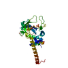
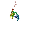
- PDB-7rm7: The X-ray crystal structure of the N-terminal domain of Staphyloc... -
+
Open data
ID or keywords:
Loading...
-
Basic information
Entry
Database: PDB / ID: 7rm7
Title
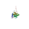
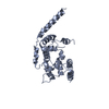
The X-ray crystal structure of the N-terminal domain of Staphylococcus aureus Fatty Acid Kinase A (FakA, residues 1-208) in complex with ADP to 1.025 Angstrom resolution
Protocol: SINGLE WAVELENGTH / Monochromatic (M) / Laue (L): M / Scattering type: x-ray
Radiation wavelength
Wavelength: 1 Å / Relative weight: 1
Reflection
Resolution: 1.025→34.24 Å / Num. obs: 95789 / % possible obs: 95.5 % / Redundancy: 6.8 % / Biso Wilson estimate: 8.3 Å2 / CC1/2: 0.999 / Rpim(I) all: 0.02 / Net I/σ(I): 19.6
Reflection shell
Resolution: 1.025→1.04 Å / Mean I/σ(I) obs: 2 / Num. unique obs: 3447 / CC1/2: 0.65 / Rpim(I) all: 0.396 / % possible all: 70.5
-
Processing
Software
Name
Version
Classification
REFMAC
5.8.0267
refinement
XDS
datareduction
Aimless
datascaling
SHELXDE
phasing
Refinement
Method to determine structure: SAD / Resolution: 1.025→34.24 Å / Cor.coef. Fo:Fc: 0.985 / Cor.coef. Fo:Fc free: 0.978 / SU B: 0.911 / SU ML: 0.02 / Cross valid method: FREE R-VALUE / ESU R: 0.022 / ESU R Free: 0.024 Details: Phasing was performed using this data set as a remote wavelength coupled with the SAD data from a cobalt-derivatized crystal (PDB ID 7RZK). Hydrogens have been used if present in the input file
Rfactor
Num. reflection
% reflection
Rfree
0.1441
4782
4.996 %
Rwork
0.1177
90938
-
all
0.119
-
-
obs
-
95720
95.158 %
Solvent computation
Ion probe radii: 0.8 Å / Shrinkage radii: 0.8 Å / VDW probe radii: 1.2 Å / Solvent model: MASK BULK SOLVENT
Displacement parameters
Biso mean: 16.035 Å2
Baniso -1
Baniso -2
Baniso -3
1-
-1.134 Å2
-0 Å2
-0 Å2
2-
-
-0.38 Å2
0 Å2
3-
-
-
1.514 Å2
Refinement step
Cycle: LAST / Resolution: 1.025→34.24 Å
Protein
Nucleic acid
Ligand
Solvent
Total
Num. atoms
1659
0
38
361
2058
Refine LS restraints
Refine-ID
Type
Dev ideal
Dev ideal target
Number
X-RAY DIFFRACTION
r_bond_refined_d
0.017
0.012
1906
X-RAY DIFFRACTION
r_bond_other_d
0.001
0.017
1814
X-RAY DIFFRACTION
r_angle_refined_deg
2.046
1.623
2605
X-RAY DIFFRACTION
r_angle_other_deg
0.67
1.565
4219
X-RAY DIFFRACTION
r_dihedral_angle_1_deg
5.361
5
265
X-RAY DIFFRACTION
r_dihedral_angle_2_deg
37.682
26.173
81
X-RAY DIFFRACTION
r_dihedral_angle_3_deg
11.704
15
349
X-RAY DIFFRACTION
r_dihedral_angle_4_deg
28.729
15
4
X-RAY DIFFRACTION
r_chiral_restr
0.103
0.2
251
X-RAY DIFFRACTION
r_gen_planes_refined
0.013
0.02
2298
X-RAY DIFFRACTION
r_gen_planes_other
0.002
0.02
393
X-RAY DIFFRACTION
r_nbd_refined
0.274
0.2
495
X-RAY DIFFRACTION
r_symmetry_nbd_other
0.192
0.2
1701
X-RAY DIFFRACTION
r_nbtor_refined
0.182
0.2
1000
X-RAY DIFFRACTION
r_symmetry_nbtor_other
0.07
0.2
926
X-RAY DIFFRACTION
r_xyhbond_nbd_refined
0.258
0.2
229
X-RAY DIFFRACTION
r_metal_ion_refined
0.092
0.2
7
X-RAY DIFFRACTION
r_symmetry_nbd_refined
0.43
0.2
30
X-RAY DIFFRACTION
r_nbd_other
0.292
0.2
96
X-RAY DIFFRACTION
r_symmetry_xyhbond_nbd_refined
0.29
0.2
53
X-RAY DIFFRACTION
r_mcbond_it
5.568
1.151
1000
X-RAY DIFFRACTION
r_mcbond_other
5.569
1.149
999
X-RAY DIFFRACTION
r_mcangle_it
6.157
1.707
1285
X-RAY DIFFRACTION
r_mcangle_other
6.155
1.708
1286
X-RAY DIFFRACTION
r_scbond_it
6.263
1.532
906
X-RAY DIFFRACTION
r_scbond_other
6.263
1.532
906
X-RAY DIFFRACTION
r_scangle_it
7.493
2.156
1320
X-RAY DIFFRACTION
r_scangle_other
7.49
2.156
1321
X-RAY DIFFRACTION
r_lrange_it
8.631
17.912
2424
X-RAY DIFFRACTION
r_lrange_other
8.007
15.916
2319
X-RAY DIFFRACTION
r_rigid_bond_restr
14.257
3
3720
LS refinement shell
Resolution (Å)
Rfactor Rfree
Num. reflection Rfree
Rfactor Rwork
Num. reflection Rwork
Refine-ID
% reflection obs (%)
1.025-1.052
0.312
261
0.293
5081
X-RAY DIFFRACTION
72.6407
1.052-1.08
0.229
289
0.214
6117
X-RAY DIFFRACTION
89.2325
1.08-1.112
0.167
333
0.157
6335
X-RAY DIFFRACTION
95.6671
1.112-1.146
0.151
335
0.141
6164
X-RAY DIFFRACTION
96.0396
1.146-1.183
0.153
312
0.134
6055
X-RAY DIFFRACTION
96.6601
1.183-1.225
0.129
329
0.123
5841
X-RAY DIFFRACTION
96.7995
1.225-1.271
0.159
301
0.122
5677
X-RAY DIFFRACTION
97.3774
1.271-1.323
0.148
292
0.117
5501
X-RAY DIFFRACTION
97.5417
1.323-1.382
0.129
274
0.115
5299
X-RAY DIFFRACTION
97.9782
1.382-1.449
0.151
262
0.111
5078
X-RAY DIFFRACTION
98.2882
1.449-1.528
0.134
242
0.106
4900
X-RAY DIFFRACTION
98.4303
1.528-1.62
0.13
241
0.099
4572
X-RAY DIFFRACTION
98.708
1.62-1.732
0.135
218
0.1
4398
X-RAY DIFFRACTION
98.9496
1.732-1.87
0.136
212
0.109
4077
X-RAY DIFFRACTION
98.9845
1.87-2.049
0.122
233
0.097
3744
X-RAY DIFFRACTION
99.3753
2.049-2.29
0.118
176
0.094
3414
X-RAY DIFFRACTION
99.281
2.29-2.643
0.122
156
0.095
3063
X-RAY DIFFRACTION
99.5054
2.643-3.234
0.14
160
0.106
2565
X-RAY DIFFRACTION
99.163
3.234-4.563
0.143
93
0.112
2012
X-RAY DIFFRACTION
96.7371
4.563-34.24
0.254
63
0.215
1045
X-RAY DIFFRACTION
86.1586
Refinement TLS params.
Method: refined / Origin x: 14.9801 Å / Origin y: 5.2362 Å / Origin z: -12.5135 Å
11
12
13
21
22
23
31
32
33
T
0.0196 Å2
0.0006 Å2
-0.0002 Å2
-
0.0153 Å2
-0.0009 Å2
-
-
0.0005 Å2
L
0.0655 °2
0.0079 °2
0.02 °2
-
0.0374 °2
-0.0074 °2
-
-
0.0965 °2
S
0.0009 Å °
0.005 Å °
-0.0057 Å °
-0.0006 Å °
0.0014 Å °
0.0002 Å °
0.0025 Å °
0.0033 Å °
-0.0023 Å °
Refinement TLS group
Selection: ALL
+
About Yorodumi
-
News
-
Feb 9, 2022. New format data for meta-information of EMDB entries
New format data for meta-information of EMDB entries
Version 3 of the EMDB header file is now the official format.
The previous official version 1.9 will be removed from the archive.
In the structure databanks used in Yorodumi, some data are registered as the other names, "COVID-19 virus" and "2019-nCoV". Here are the details of the virus and the list of structure data.
Jan 31, 2019. EMDB accession codes are about to change! (news from PDBe EMDB page)
EMDB accession codes are about to change! (news from PDBe EMDB page)
The allocation of 4 digits for EMDB accession codes will soon come to an end. Whilst these codes will remain in use, new EMDB accession codes will include an additional digit and will expand incrementally as the available range of codes is exhausted. The current 4-digit format prefixed with “EMD-” (i.e. EMD-XXXX) will advance to a 5-digit format (i.e. EMD-XXXXX), and so on. It is currently estimated that the 4-digit codes will be depleted around Spring 2019, at which point the 5-digit format will come into force.
The EM Navigator/Yorodumi systems omit the EMD- prefix.
Related info.:Q: What is EMD? / ID/Accession-code notation in Yorodumi/EM Navigator
Yorodumi is a browser for structure data from EMDB, PDB, SASBDB, etc.
This page is also the successor to EM Navigator detail page, and also detail information page/front-end page for Omokage search.
The word "yorodu" (or yorozu) is an old Japanese word meaning "ten thousand". "mi" (miru) is to see.
Related info.:EMDB / PDB / SASBDB / Comparison of 3 databanks / Yorodumi Search / Aug 31, 2016. New EM Navigator & Yorodumi / Yorodumi Papers / Jmol/JSmol / Function and homology information / Changes in new EM Navigator and Yorodumi
 Movie
Movie Controller

 Yorodumi
 Open data
 Basic information
 Components
 Keywords
 Function and homology information
 Staphylococcus aureus (bacteria)
 X-RAY DIFFRACTION /
  Authors
 Citation
 Structure visualization
 Downloads & links
 Other downloads


 PDBj
PDBj Assembly
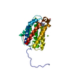

 Mass: 92.094 Da / Num. of mol.: 1 / Source method: obtained synthetically / Formula: C3H8O3
 Mass: 427.201 Da / Num. of mol.: 1 / Source method: obtained synthetically / Formula: C10H15N5O10P2 / Feature type: SUBJECT OF INVESTIGATION / Comment: ADP, energy-carrying molecule*YM
 Mass: 24.305 Da / Num. of mol.: 5 / Source method: obtained synthetically / Formula: Mg / Feature type: SUBJECT OF INVESTIGATION
 Mass: 18.015 Da / Num. of mol.: 361 / Source method: isolated from a natural source / Formula: H2O
 Sample preparation
 / Beamline: 22-ID / Wavelength: 1 Å
 Processing