 Movie
Movie Controller


+ Open data

- Basic information
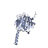
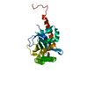
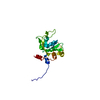
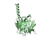
| Entry | Database: PDB / ID: 2dyt | ||||||
|---|---|---|---|---|---|---|---|
| Title | The crystal structure of Saccharomyces cerevisiae Atg3 | ||||||
 Components Components | Autophagy-related protein 3 | ||||||
 Keywords Keywords | LIGASE / E2 fold | ||||||
| Function / homology |  Function and homology information Function and homology informationAtg8-family conjugating enzyme activity / Atg8-family ligase activity / phagophore / Macroautophagy / glycophagy / cytoplasm to vacuole targeting by the Cvt pathway / nucleophagy / autophagy of mitochondrion / piecemeal microautophagy of the nucleus / phagophore assembly site ...Atg8-family conjugating enzyme activity / Atg8-family ligase activity / phagophore / Macroautophagy / glycophagy / cytoplasm to vacuole targeting by the Cvt pathway / nucleophagy / autophagy of mitochondrion / piecemeal microautophagy of the nucleus / phagophore assembly site / protein targeting to membrane / autophagosome assembly / mitochondrion / cytosol / cytoplasm Similarity search - Function | ||||||
| Biological species |  | ||||||
| Method |  X-RAY DIFFRACTION / SYNCHROTRON / MIR / Resolution: 2.5 Å X-RAY DIFFRACTION / SYNCHROTRON / MIR / Resolution: 2.5 Å | ||||||
 Authors Authors | Yamada, Y. / Suzuki, N.N. / Inagaki, F. | ||||||
 Citation Citation | Journal: J.Biol.Chem. / Year: 2007 Title: The crystal structure of Atg3, an autophagy-related ubiquitin carrier protein (E2) enzyme that mediates Atg8 lipidation Authors: Yamada, Y. / Suzuki, N.N. / Hanada, T. / Ichimura, Y. / Kumeta, H. / Fujioka, Y. / Ohsumi, Y. / Inagaki, F. #1: Journal: ACTA CRYSTALLOGR.,SECT.F / Year: 2006 Title: Crystallization and preliminary X-ray analysis of Atg3 Authors: Yamada, Y. / Suzuki, N.N. / Fujioka, Y. / Ichimura, Y. / Ohsumi, Y. / Inagaki, F. | ||||||
| History |
|
- Structure visualization
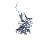
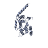
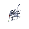
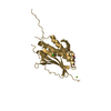
| Structure viewer | Molecule: MolmilJmol/JSmol |
|---|
- Downloads & links
-Download
| PDBx/mmCIF format | 2dyt.cif.gz | 58.2 KB | Display | PDBx/mmCIF format |
|---|---|---|---|---|
| PDB format | pdb2dyt.ent.gz | 40.5 KB | Display | PDB format |
| PDBx/mmJSON format | 2dyt.json.gz | Tree view | PDBx/mmJSON format | |
| Others |  Other downloads Other downloads |
-Validation report
| Arichive directory | https://data.pdbj.org/pub/pdb/validation_reports/dy/2dytftp://data.pdbj.org/pub/pdb/validation_reports/dy/2dyt | HTTPS FTP |
|---|
-Related structure data
| Similar structure data |
|---|
-Links
 PDBj
PDBj
- Assembly
| Deposited unit | 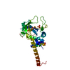
| ||||||||
|---|---|---|---|---|---|---|---|---|---|
| 1 |
| ||||||||
| Unit cell |
|
-Components
| #1: Protein | Mass: 36071.168 Da / Num. of mol.: 1 Source method: isolated from a genetically manipulated source Source: (gene. exp.) Production host:  | ||
|---|---|---|---|
| #2: Chemical |   Mass: 96.063 Da / Num. of mol.: 2 / Source method: obtained synthetically / Formula: SO4 Mass: 96.063 Da / Num. of mol.: 2 / Source method: obtained synthetically / Formula: SO4#3: Water | ChemComp-HOH / |  Mass: 18.015 Da / Num. of mol.: 46 / Source method: isolated from a natural source / Formula: H2O Mass: 18.015 Da / Num. of mol.: 46 / Source method: isolated from a natural source / Formula: H2O |
-Experimental details
-Experiment
| Experiment | Method: X-RAY DIFFRACTION / Number of used crystals: 1 |
|---|
- Sample preparation
| Crystal | Density Matthews: 2.79 Å3/Da / Density % sol: 55.95 % |
|---|---|
| Crystal grow | Temperature: 293 K / Method: vapor diffusion, sitting drop / pH: 5.8 Details: 0.3M ammonium sulfate, 1.0M lithium sulfate, 0.1M citrate, pH 5.8, VAPOR DIFFUSION, SITTING DROP, temperature 293K |
-Data collection
| Diffraction | Mean temperature: 90 K |
|---|---|
| Diffraction source | Source: SYNCHROTRON / Site: SPring-8  / Beamline: BL41XU / Wavelength: 1 Å / Beamline: BL41XU / Wavelength: 1 Å |
| Detector | Type: ADSC QUANTUM 315 / Detector: CCD |
| Radiation | Protocol: SINGLE WAVELENGTH / Monochromatic (M) / Laue (L): M / Scattering type: x-ray |
| Radiation wavelength | Wavelength: 1 Å / Relative weight: 1 |
| Reflection | Resolution: 2.5→50 Å / Num. all: 83016 / Num. obs: 83016 / % possible obs: 98.8 % / Observed criterion σ(F): 0 / Observed criterion σ(I): -3 / Redundancy: 6.1 % / Biso Wilson estimate: 51.3 Å2 / Rmerge(I) obs: 0.06 / Net I/σ(I): 22.3 |
| Reflection shell | Resolution: 2.5→2.6 Å / Rmerge(I) obs: 0.271 / % possible all: 99.6 |
- Processing
| Software |
| ||||||||||||||||||||||||||||||||||||||||||||||||||||||||||||||||||||||||||||||||
|---|---|---|---|---|---|---|---|---|---|---|---|---|---|---|---|---|---|---|---|---|---|---|---|---|---|---|---|---|---|---|---|---|---|---|---|---|---|---|---|---|---|---|---|---|---|---|---|---|---|---|---|---|---|---|---|---|---|---|---|---|---|---|---|---|---|---|---|---|---|---|---|---|---|---|---|---|---|---|---|---|---|
| Refinement | Method to determine structure: MIR / Resolution: 2.5→33.84 Å / Rfactor Rfree error: 0.007 / Data cutoff high absF: 756551.72 / Data cutoff low absF: 0 / Isotropic thermal model: RESTRAINED / Cross valid method: THROUGHOUT / σ(F): 0 / σ(I): -3 / Stereochemistry target values: Engh & Huber
| ||||||||||||||||||||||||||||||||||||||||||||||||||||||||||||||||||||||||||||||||
| Solvent computation | Solvent model: FLAT MODEL / Bsol: 60.775 Å2 / ksol: 0.37149 e/Å3 | ||||||||||||||||||||||||||||||||||||||||||||||||||||||||||||||||||||||||||||||||
| Displacement parameters | Biso mean: 54.2 Å2
| ||||||||||||||||||||||||||||||||||||||||||||||||||||||||||||||||||||||||||||||||
| Refine analyze |
| ||||||||||||||||||||||||||||||||||||||||||||||||||||||||||||||||||||||||||||||||
| Refinement step | Cycle: LAST / Resolution: 2.5→33.84 Å
| ||||||||||||||||||||||||||||||||||||||||||||||||||||||||||||||||||||||||||||||||
| Refine LS restraints |
| ||||||||||||||||||||||||||||||||||||||||||||||||||||||||||||||||||||||||||||||||
| LS refinement shell | Resolution: 2.5→2.66 Å / Rfactor Rfree error: 0.022 / Total num. of bins used: 6
| ||||||||||||||||||||||||||||||||||||||||||||||||||||||||||||||||||||||||||||||||
| Xplor file |
|
