 Movie
Movie Controller

[English] 日本語
 Yorodumi
- PDB-7rfl: CamA Adenine Methyltransferase Complexed to Cognate Substrate DNA... -
+ Open data

- Basic information
| Entry | Database: PDB / ID: 7rfl | ||||||
|---|---|---|---|---|---|---|---|
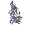
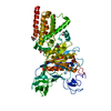
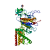
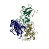
| Title | CamA Adenine Methyltransferase Complexed to Cognate Substrate DNA and Inhibitor SGC0946 | ||||||
 Components Components |
| ||||||
 Keywords Keywords | DNA BINDING PROTEIN/DNA / DNA Adenine Methylation / PROTEIN-DNA COMPLEX / TRANSFERASE / DNA BINDING PROTEIN / DNA BINDING PROTEIN-DNA complex | ||||||
| Function / homology |  Function and homology information Function and homology informationsite-specific DNA-methyltransferase (adenine-specific) / site-specific DNA-methyltransferase (adenine-specific) activity / DNA restriction-modification system / methylation / DNA binding Similarity search - Function | ||||||
| Biological species |  Clostridioides difficile (bacteria) Clostridioides difficile (bacteria) | ||||||
| Method |  X-RAY DIFFRACTION / SYNCHROTRON / MOLECULAR REPLACEMENT / Resolution: 2.38 Å X-RAY DIFFRACTION / SYNCHROTRON / MOLECULAR REPLACEMENT / Resolution: 2.38 Å | ||||||
 Authors Authors | Horton, J.R. / Cheng, X. / Zhou, J. | ||||||
| Funding support |  United States, 1items United States, 1items
| ||||||
 Citation Citation | Journal: Epigenetics / Year: 2022 Title: Repurposing epigenetic inhibitors to target the Clostridioides difficile- specific DNA adenine methyltransferase and sporulation regulator CamA. Authors: Zhou, J. / Horton, J.R. / Yu, D. / Ren, R. / Blumenthal, R.M. / Zhang, X. / Cheng, X. | ||||||
| History |
|
- Structure visualization
| Structure viewer | Molecule: MolmilJmol/JSmol |
|---|
- Downloads & links
-Download
| PDBx/mmCIF format | 7rfl.cif.gz | 467.7 KB | Display | PDBx/mmCIF format |
|---|---|---|---|---|
| PDB format | pdb7rfl.ent.gz | 318.5 KB | Display | PDB format |
| PDBx/mmJSON format | 7rfl.json.gz | Tree view | PDBx/mmJSON format | |
| Others |  Other downloads Other downloads |
-Validation report
| Arichive directory | https://data.pdbj.org/pub/pdb/validation_reports/rf/7rflftp://data.pdbj.org/pub/pdb/validation_reports/rf/7rfl | HTTPS FTP |
|---|
-Related structure data
| Related structure data | 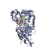 7rfkC  7rfmC 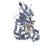 7rfnC 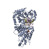 7lt5S S: Starting model for refinement C: citing same article ( |
|---|---|
| Similar structure data |


-Links
 PDBj
PDBj


- Assembly
| Deposited unit | 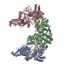
| ||||||||||||
|---|---|---|---|---|---|---|---|---|---|---|---|---|---|
| 1 | 
| ||||||||||||
| 2 | 
| ||||||||||||
| 3 | 
| ||||||||||||
| Unit cell |
|
-Components
-Protein , 1 types, 3 molecules ABC
| #1: Protein | Mass: 68887.172 Da / Num. of mol.: 3 Source method: isolated from a genetically manipulated source Source: (gene. exp.) Clostridioides difficile (bacteria) / Strain: 630 / Gene: CD630_27580 / Production host: References: UniProt: Q183J3, site-specific DNA-methyltransferase (adenine-specific) |
|---|
-DNA chain , 2 types, 6 molecules EGIDFH
| #2: DNA chain | Mass: 4325.825 Da / Num. of mol.: 3 / Source method: obtained synthetically / Source: (synth.) Clostridioides difficile (bacteria)#3: DNA chain | Mass: 4232.795 Da / Num. of mol.: 3 / Source method: obtained synthetically / Source: (synth.) Clostridioides difficile (bacteria) |
|---|
-Non-polymers , 4 types, 455 molecules 


| #4: Chemical |  Mass: 618.566 Da / Num. of mol.: 3 / Source method: obtained synthetically / Formula: C28H40BrN7O4 / Feature type: SUBJECT OF INVESTIGATION Mass: 618.566 Da / Num. of mol.: 3 / Source method: obtained synthetically / Formula: C28H40BrN7O4 / Feature type: SUBJECT OF INVESTIGATION#5: Chemical | ChemComp-K /  Mass: 39.098 Da / Num. of mol.: 5 / Source method: obtained synthetically / Formula: K Mass: 39.098 Da / Num. of mol.: 5 / Source method: obtained synthetically / Formula: K#6: Chemical | ChemComp-EDO /  Mass: 62.068 Da / Num. of mol.: 8 / Source method: obtained synthetically / Formula: C2H6O2 Mass: 62.068 Da / Num. of mol.: 8 / Source method: obtained synthetically / Formula: C2H6O2#7: Water | ChemComp-HOH / | Mass: 18.015 Da / Num. of mol.: 439 / Source method: isolated from a natural source / Formula: H2O |
|---|
-Details
| Has ligand of interest | Y |
|---|
-Experimental details
-Experiment
| Experiment | Method: X-RAY DIFFRACTION / Number of used crystals: 1 |
|---|
- Sample preparation
| Crystal | Density Matthews: 3.26 Å3/Da / Density % sol: 62.25 % |
|---|---|
| Crystal grow | Temperature: 292 K / Method: vapor diffusion, sitting drop Details: 21~24% (w/v) polyethylene glycol 3350, 0.1 M Tris-HCl pH 7.0~7.5, 0.28 M potassium citrate PH range: 7-7.5 |
-Data collection
| Diffraction | Mean temperature: 100 K / Serial crystal experiment: N |
|---|---|
| Diffraction source | Source: SYNCHROTRON / Site: APS / Beamline: 22-ID / Wavelength: 1 Å |
| Detector | Type: DECTRIS EIGER X 16M / Detector: PIXEL / Date: Feb 7, 2021 |
| Radiation | Protocol: SINGLE WAVELENGTH / Monochromatic (M) / Laue (L): M / Scattering type: x-ray |
| Radiation wavelength | Wavelength: 1 Å / Relative weight: 1 |
| Reflection | Resolution: 2.38→41.87 Å / Num. obs: 111448 / % possible obs: 91.6 % / Redundancy: 10.8 % / Biso Wilson estimate: 44.91 Å2 / Rmerge(I) obs: 0.206 / Rpim(I) all: 0.062 / Net I/σ(I): 10.4 |
| Reflection shell | Resolution: 2.38→2.48 Å / Rmerge(I) obs: 1.78 / Mean I/σ(I) obs: 1.5 / Num. unique obs: 9690 / CC1/2: 0.258 / Rpim(I) all: 0.615 / % possible all: 80.4 |
- Processing
| Software |
| |||||||||||||||||||||||||||||||||||||||||||||||||||||||||||||||||||||||||||||||||||||||||||||||||||||||||
|---|---|---|---|---|---|---|---|---|---|---|---|---|---|---|---|---|---|---|---|---|---|---|---|---|---|---|---|---|---|---|---|---|---|---|---|---|---|---|---|---|---|---|---|---|---|---|---|---|---|---|---|---|---|---|---|---|---|---|---|---|---|---|---|---|---|---|---|---|---|---|---|---|---|---|---|---|---|---|---|---|---|---|---|---|---|---|---|---|---|---|---|---|---|---|---|---|---|---|---|---|---|---|---|---|---|---|
| Refinement | Method to determine structure: MOLECULAR REPLACEMENT Starting model: 7LT5 Resolution: 2.38→41.87 Å / SU ML: 0.2826 / Cross valid method: FREE R-VALUE / σ(F): 1.34 / Phase error: 25.9711 Stereochemistry target values: GeoStd + Monomer Library + CDL v1.2
| |||||||||||||||||||||||||||||||||||||||||||||||||||||||||||||||||||||||||||||||||||||||||||||||||||||||||
| Solvent computation | Shrinkage radii: 0.9 Å / VDW probe radii: 1.11 Å / Solvent model: FLAT BULK SOLVENT MODEL | |||||||||||||||||||||||||||||||||||||||||||||||||||||||||||||||||||||||||||||||||||||||||||||||||||||||||
| Displacement parameters | Biso mean: 56.33 Å2 | |||||||||||||||||||||||||||||||||||||||||||||||||||||||||||||||||||||||||||||||||||||||||||||||||||||||||
| Refinement step | Cycle: LAST / Resolution: 2.38→41.87 Å
| |||||||||||||||||||||||||||||||||||||||||||||||||||||||||||||||||||||||||||||||||||||||||||||||||||||||||
| Refine LS restraints |
| |||||||||||||||||||||||||||||||||||||||||||||||||||||||||||||||||||||||||||||||||||||||||||||||||||||||||
| LS refinement shell |
|
