 Movie
Movie Controller

[English] 日本語
 Yorodumi
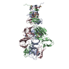
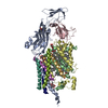
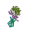
- PDB-7rej: Tailspike protein 4 (TSP4) from phage CBA120, residues 1-335, obt... -
+ Open data

- Basic information
| Entry | Database: PDB / ID: 7rej | ||||||
|---|---|---|---|---|---|---|---|
| Title | Tailspike protein 4 (TSP4) from phage CBA120, residues 1-335, obtained in the presence of NaK-Tartrate | ||||||
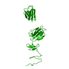
 Components Components | Tailspike protein | ||||||
 Keywords Keywords | VIRAL PROTEIN / tailspike protein-protein interaction / TSP4 attachment to the tail baseplate / triple beta-helix / beta jellyroll | ||||||
| Function / homology | Tail spike TSP1/Gp66, N-terminal domain / Tail spike TSP1/Gp66 receptor binding N-terminal domain / IMIDAZOLE / Tailspike protein Function and homology information Function and homology information | ||||||
| Biological species |  Escherichia virus CBA120 Escherichia virus CBA120 | ||||||
| Method |  X-RAY DIFFRACTION / SYNCHROTRON / MOLECULAR REPLACEMENT / Resolution: 2.6 Å X-RAY DIFFRACTION / SYNCHROTRON / MOLECULAR REPLACEMENT / Resolution: 2.6 Å | ||||||
 Authors Authors | Chao, K. / Shang, X. / Grenfield, J. / Linden, S.B. / Nelson, D.C. / Herzberg, O. | ||||||
| Funding support |  United States, 1items United States, 1items
| ||||||
 Citation Citation | Journal: Sci Rep / Year: 2022 Title: Structure of Escherichia coli O157:H7 bacteriophage CBA120 tailspike protein 4 baseplate anchor and tailspike assembly domains (TSP4-N). Authors: Chao, K.L. / Shang, X. / Greenfield, J. / Linden, S.B. / Alreja, A.B. / Nelson, D.C. / Herzberg, O. | ||||||
| History |
|
- Structure visualization
| Structure viewer | Molecule: MolmilJmol/JSmol |
|---|
- Downloads & links
-Download
| PDBx/mmCIF format | 7rej.cif.gz | 229.9 KB | Display | PDBx/mmCIF format |
|---|---|---|---|---|
| PDB format | pdb7rej.ent.gz | 186.9 KB | Display | PDB format |
| PDBx/mmJSON format | 7rej.json.gz | Tree view | PDBx/mmJSON format | |
| Others |  Other downloads Other downloads |
-Validation report
| Arichive directory | https://data.pdbj.org/pub/pdb/validation_reports/re/7rejftp://data.pdbj.org/pub/pdb/validation_reports/re/7rej | HTTPS FTP |
|---|
-Related structure data


-Links
 PDBj
PDBj


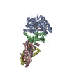
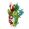
- Assembly
| Deposited unit | 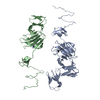
| |||||||||||||||||||||
|---|---|---|---|---|---|---|---|---|---|---|---|---|---|---|---|---|---|---|---|---|---|---|
| 1 | 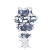
| |||||||||||||||||||||
| 2 | 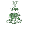
| |||||||||||||||||||||
| Unit cell |
| |||||||||||||||||||||
| Components on special symmetry positions |
|
-Components
| #1: Protein | Mass: 36320.750 Da / Num. of mol.: 2 / Fragment: N-terminal domain (UNP residues 1-335) Source method: isolated from a genetically manipulated source Source: (gene. exp.) Escherichia virus CBA120 / Gene: orf213 / Production host:  #2: Chemical |   Mass: 69.085 Da / Num. of mol.: 2 / Source method: obtained synthetically / Formula: C3H5N2 Mass: 69.085 Da / Num. of mol.: 2 / Source method: obtained synthetically / Formula: C3H5N2#3: Water | ChemComp-HOH / |  Mass: 18.015 Da / Num. of mol.: 31 / Source method: isolated from a natural source / Formula: H2O Mass: 18.015 Da / Num. of mol.: 31 / Source method: isolated from a natural source / Formula: H2OHas ligand of interest | N | |
|---|
-Experimental details
-Experiment
| Experiment | Method: X-RAY DIFFRACTION / Number of used crystals: 1 |
|---|
- Sample preparation
| Crystal | Density Matthews: 2.64 Å3/Da / Density % sol: 53.34 % |
|---|---|
| Crystal grow | Temperature: 295 K / Method: vapor diffusion / pH: 7.8 Details: 1 M potassium sodium tartrate, 0.2 M sodium chloride, 0.1 M imidazole, pH 7.8 |
-Data collection
| Diffraction | Mean temperature: 100 K / Serial crystal experiment: N |
|---|---|
| Diffraction source | Source: SYNCHROTRON / Site: APS / Beamline: 23-ID-B / Wavelength: 1.0332 Å |
| Detector | Type: DECTRIS EIGER X 16M / Detector: PIXEL / Date: Jul 3, 2018 / Details: mirrors |
| Radiation | Monochromator: double crystal monochromato / Protocol: SINGLE WAVELENGTH / Monochromatic (M) / Laue (L): M / Scattering type: x-ray |
| Radiation wavelength | Wavelength: 1.0332 Å / Relative weight: 1 |
| Reflection | Resolution: 2.5→27.37 Å / Num. obs: 25427 / % possible obs: 98.9 % / Redundancy: 2.6 % / Biso Wilson estimate: 52 Å2 / CC1/2: 0.987 / Rmerge(I) obs: 0.09 / Net I/σ(I): 5.1 |
| Reflection shell | Resolution: 2.5→2.6 Å / Rmerge(I) obs: 0.85 / Num. unique obs: 2897 / % possible all: 98.8 |
- Processing
| Software |
| ||||||||||||||||||||||||||||||||||||||||||||||||||||||||||||||||||||||||||||||||||||||||||||||||||||||||||||||||||||||||||||||||||||||||||||||||||||||
|---|---|---|---|---|---|---|---|---|---|---|---|---|---|---|---|---|---|---|---|---|---|---|---|---|---|---|---|---|---|---|---|---|---|---|---|---|---|---|---|---|---|---|---|---|---|---|---|---|---|---|---|---|---|---|---|---|---|---|---|---|---|---|---|---|---|---|---|---|---|---|---|---|---|---|---|---|---|---|---|---|---|---|---|---|---|---|---|---|---|---|---|---|---|---|---|---|---|---|---|---|---|---|---|---|---|---|---|---|---|---|---|---|---|---|---|---|---|---|---|---|---|---|---|---|---|---|---|---|---|---|---|---|---|---|---|---|---|---|---|---|---|---|---|---|---|---|---|---|---|---|---|
| Refinement | Method to determine structure: MOLECULAR REPLACEMENT Starting model: SeMet TSP4-N(1-335) structure Resolution: 2.6→20 Å / Cor.coef. Fo:Fc: 0.949 / Cor.coef. Fo:Fc free: 0.932 / SU B: 26.051 / SU ML: 0.244 / Cross valid method: THROUGHOUT / σ(F): 0 / ESU R: 0.554 / ESU R Free: 0.274 / Stereochemistry target values: MAXIMUM LIKELIHOOD Details: U VALUES : WITH TLS ADDED HYDROGENS HAVE BEEN ADDED IN THE RIDING POSITIONS
| ||||||||||||||||||||||||||||||||||||||||||||||||||||||||||||||||||||||||||||||||||||||||||||||||||||||||||||||||||||||||||||||||||||||||||||||||||||||
| Solvent computation | Ion probe radii: 0.8 Å / Shrinkage radii: 0.8 Å / VDW probe radii: 1.2 Å / Solvent model: MASK | ||||||||||||||||||||||||||||||||||||||||||||||||||||||||||||||||||||||||||||||||||||||||||||||||||||||||||||||||||||||||||||||||||||||||||||||||||||||
| Displacement parameters | Biso max: 193.16 Å2 / Biso mean: 80.295 Å2 / Biso min: 29.86 Å2
| ||||||||||||||||||||||||||||||||||||||||||||||||||||||||||||||||||||||||||||||||||||||||||||||||||||||||||||||||||||||||||||||||||||||||||||||||||||||
| Refinement step | Cycle: final / Resolution: 2.6→20 Å
| ||||||||||||||||||||||||||||||||||||||||||||||||||||||||||||||||||||||||||||||||||||||||||||||||||||||||||||||||||||||||||||||||||||||||||||||||||||||
| Refine LS restraints |
| ||||||||||||||||||||||||||||||||||||||||||||||||||||||||||||||||||||||||||||||||||||||||||||||||||||||||||||||||||||||||||||||||||||||||||||||||||||||
| LS refinement shell | Resolution: 2.6→2.666 Å / Rfactor Rfree error: 0 / Total num. of bins used: 20
| ||||||||||||||||||||||||||||||||||||||||||||||||||||||||||||||||||||||||||||||||||||||||||||||||||||||||||||||||||||||||||||||||||||||||||||||||||||||
| Refinement TLS params. | Method: refined / Refine-ID: X-RAY DIFFRACTION
| ||||||||||||||||||||||||||||||||||||||||||||||||||||||||||||||||||||||||||||||||||||||||||||||||||||||||||||||||||||||||||||||||||||||||||||||||||||||
| Refinement TLS group |
|
