 Movie
Movie Controller

[English] 日本語
 Yorodumi
- PDB-1nen: Complex II (Succinate Dehydrogenase) From E. Coli with Dinitrophe... -
+ Open data

- Basic information
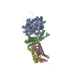
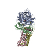
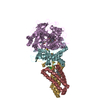
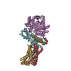
| Entry | Database: PDB / ID: 1nen | ||||||
|---|---|---|---|---|---|---|---|
| Title | Complex II (Succinate Dehydrogenase) From E. Coli with Dinitrophenol-17 inhibitor co-crystallized at the ubiquinone binding site | ||||||
 Components Components | (Succinate dehydrogenase ...) x 4 | ||||||
 Keywords Keywords | OXIDOREDUCTASE/ELECTRON TRANSPORT / membrane protein / respiratory complex / OXIDOREDUCTASE-ELECTRON TRANSPORT COMPLEX | ||||||
| Function / homology |  Function and homology information Function and homology informationsuccinate dehydrogenase activity / respiratory chain complex II (succinate dehydrogenase) / succinate dehydrogenase (quinone) activity / succinate dehydrogenase / cytochrome complex assembly / aerobic electron transport chain / anaerobic respiration / 3 iron, 4 sulfur cluster binding / ubiquinone binding / iron-sulfur cluster binding ...succinate dehydrogenase activity / respiratory chain complex II (succinate dehydrogenase) / succinate dehydrogenase (quinone) activity / succinate dehydrogenase / cytochrome complex assembly / aerobic electron transport chain / anaerobic respiration / 3 iron, 4 sulfur cluster binding / ubiquinone binding / iron-sulfur cluster binding / tricarboxylic acid cycle / aerobic respiration / respiratory electron transport chain / 2 iron, 2 sulfur cluster binding / flavin adenine dinucleotide binding / 4 iron, 4 sulfur cluster binding / electron transfer activity / heme binding / membrane / metal ion binding / plasma membrane Similarity search - Function | ||||||
| Biological species |  | ||||||
| Method |  X-RAY DIFFRACTION / SYNCHROTRON / MOLECULAR REPLACEMENT / Resolution: 2.9 Å X-RAY DIFFRACTION / SYNCHROTRON / MOLECULAR REPLACEMENT / Resolution: 2.9 Å | ||||||
 Authors Authors | Yankovskaya, V. / Horsefield, R. / Tornroth, S. / Luna-Chavez, C. / Miyoshi, H. / Leger, C. / Byrne, B. / Cecchini, G. / Iwata, S. | ||||||
 Citation Citation | Journal: Science / Year: 2003 Title: Architecture of succinate dehydrogenase and reactive oxygen species generation Authors: Yankovskaya, V. / Horsefield, R. / Tornroth, S. / Luna-Chavez, C. / Miyoshi, H. / Leger, C. / Byrne, B. / Cecchini, G. / Iwata, S. | ||||||
| History |
| ||||||
| Remark 600 | HETEROGEN THE HET GROUP CDN WAS NAMED CARDIOLIPIN WHICH IS A GENERIC NAME FOR THIS TYPE OF LIPID. ...HETEROGEN THE HET GROUP CDN WAS NAMED CARDIOLIPIN WHICH IS A GENERIC NAME FOR THIS TYPE OF LIPID. THE 4 TAILS OF THE MOLECULE ARE DISORDERED IN THE STRUCTURE AND THEIR EXACT LENGTH CAN NOT ASCERTAINED MAKING IT DIFFICULT TO ASSIGN AN EXACT NAME. |
- Structure visualization
| Structure viewer | Molecule: MolmilJmol/JSmol |
|---|
- Downloads & links
-Download
| PDBx/mmCIF format | 1nen.cif.gz | 238.7 KB | Display | PDBx/mmCIF format |
|---|---|---|---|---|
| PDB format | pdb1nen.ent.gz | 184.5 KB | Display | PDB format |
| PDBx/mmJSON format | 1nen.json.gz | Tree view | PDBx/mmJSON format | |
| Others |  Other downloads Other downloads |
-Validation report
| Arichive directory | https://data.pdbj.org/pub/pdb/validation_reports/ne/1nenftp://data.pdbj.org/pub/pdb/validation_reports/ne/1nen | HTTPS FTP |
|---|
-Related structure data


-Links
 PDBj
PDBj


- Assembly
| Deposited unit | 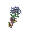
| ||||||||
|---|---|---|---|---|---|---|---|---|---|
| 1 |
| ||||||||
| 2 | 
| ||||||||
| 3 | 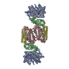
| ||||||||
| Unit cell |
|
-Components
-Succinate dehydrogenase ... , 4 types, 4 molecules ABCD
| #1: Protein | Mass: 64502.766 Da / Num. of mol.: 1 Source method: isolated from a genetically manipulated source Source: (gene. exp.) References: UniProt: P0AC41, EC: 1.3.99.1, succinate dehydrogenase |
|---|---|
| #2: Protein | Mass: 26800.912 Da / Num. of mol.: 1 Source method: isolated from a genetically manipulated source Source: (gene. exp.) References: UniProt: P07014, EC: 1.3.99.1, succinate dehydrogenase |
| #3: Protein | Mass: 14313.100 Da / Num. of mol.: 1 Source method: isolated from a genetically manipulated source Source: (gene. exp.) |
| #4: Protein | Mass: 12874.438 Da / Num. of mol.: 1 Source method: isolated from a genetically manipulated source Source: (gene. exp.) |
-Non-polymers , 11 types, 151 molecules 


| #5: Chemical | ChemComp-OAA /  Mass: 131.064 Da / Num. of mol.: 1 / Source method: obtained synthetically / Formula: C4H3O5 Mass: 131.064 Da / Num. of mol.: 1 / Source method: obtained synthetically / Formula: C4H3O5 | ||||||||||||||||||
|---|---|---|---|---|---|---|---|---|---|---|---|---|---|---|---|---|---|---|---|
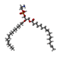
| #6: Chemical |  Mass: 40.078 Da / Num. of mol.: 2 / Source method: obtained synthetically / Formula: Ca Mass: 40.078 Da / Num. of mol.: 2 / Source method: obtained synthetically / Formula: Ca#7: Chemical | ChemComp-FAD / |  Mass: 785.550 Da / Num. of mol.: 1 / Source method: obtained synthetically / Formula: C27H33N9O15P2 / Comment: FAD*YM Mass: 785.550 Da / Num. of mol.: 1 / Source method: obtained synthetically / Formula: C27H33N9O15P2 / Comment: FAD*YM#8: Chemical | ChemComp-FES / |  Mass: 175.820 Da / Num. of mol.: 1 / Source method: obtained synthetically / Formula: Fe2S2 Mass: 175.820 Da / Num. of mol.: 1 / Source method: obtained synthetically / Formula: Fe2S2#9: Chemical | ChemComp-SF4 / |  Mass: 351.640 Da / Num. of mol.: 1 / Source method: obtained synthetically / Formula: Fe4S4 Mass: 351.640 Da / Num. of mol.: 1 / Source method: obtained synthetically / Formula: Fe4S4#10: Chemical | ChemComp-F3S / |  Mass: 295.795 Da / Num. of mol.: 1 / Source method: obtained synthetically / Formula: Fe3S4 Mass: 295.795 Da / Num. of mol.: 1 / Source method: obtained synthetically / Formula: Fe3S4#11: Chemical | ChemComp-HEM / |  Mass: 616.487 Da / Num. of mol.: 1 / Source method: obtained synthetically / Formula: C34H32FeN4O4 Mass: 616.487 Da / Num. of mol.: 1 / Source method: obtained synthetically / Formula: C34H32FeN4O4#12: Chemical | ChemComp-DNT / |  Mass: 282.292 Da / Num. of mol.: 1 / Source method: obtained synthetically / Formula: C13H18N2O5 Mass: 282.292 Da / Num. of mol.: 1 / Source method: obtained synthetically / Formula: C13H18N2O5#13: Chemical | ChemComp-CDN / |  Mass: 1151.511 Da / Num. of mol.: 1 / Source method: obtained synthetically / Formula: C58H120O17P2 Mass: 1151.511 Da / Num. of mol.: 1 / Source method: obtained synthetically / Formula: C58H120O17P2#14: Chemical | ChemComp-EPH / |  Mass: 709.933 Da / Num. of mol.: 1 / Source method: obtained synthetically / Formula: C39H68NO8P / Comment: phospholipid*YM Mass: 709.933 Da / Num. of mol.: 1 / Source method: obtained synthetically / Formula: C39H68NO8P / Comment: phospholipid*YM#15: Water | ChemComp-HOH / | Mass: 18.015 Da / Num. of mol.: 140 / Source method: isolated from a natural source / Formula: H2O |
-Details
| Has protein modification | Y |
|---|
-Experimental details
-Experiment
| Experiment | Method: X-RAY DIFFRACTION / Number of used crystals: 1 |
|---|
- Sample preparation
| Crystal | Density Matthews: 4.08 Å3/Da / Density % sol: 69.86 % | ||||||||||||||||||||||||||||||||||||||||||
|---|---|---|---|---|---|---|---|---|---|---|---|---|---|---|---|---|---|---|---|---|---|---|---|---|---|---|---|---|---|---|---|---|---|---|---|---|---|---|---|---|---|---|---|
| Crystal grow | Temperature: 293 K / Method: vapor diffusion, hanging drop / pH: 8.2 Details: Tris-HCl, CaCl2, PEG 400, BaCl2, ethylene glycol, DNP17, pH 8.2, VAPOR DIFFUSION, HANGING DROP, temperature 293.0K | ||||||||||||||||||||||||||||||||||||||||||
| Crystal grow | *PLUS Temperature: 20 ℃ | ||||||||||||||||||||||||||||||||||||||||||
| Components of the solutions | *PLUS
|
-Data collection
| Diffraction | Mean temperature: 100 K |
|---|---|
| Diffraction source | Source: SYNCHROTRON / Site: ESRF  / Beamline: ID14-2 / Wavelength: 0.915 Å / Beamline: ID14-2 / Wavelength: 0.915 Å |
| Detector | Type: ADSC QUANTUM 4 / Detector: CCD / Date: Jul 7, 2002 |
| Radiation | Protocol: SINGLE WAVELENGTH / Monochromatic (M) / Laue (L): M / Scattering type: x-ray |
| Radiation wavelength | Wavelength: 0.915 Å / Relative weight: 1 |
| Reflection | Resolution: 2.9→40 Å / Num. all: 43562 / Num. obs: 42735 / % possible obs: 98.1 % / Observed criterion σ(F): 0 / Observed criterion σ(I): -3 / Redundancy: 2.8 % / Rsym value: 0.116 |
| Reflection shell | Resolution: 2.9→2.96 Å / Rsym value: 0.428 / % possible all: 96.6 |
| Reflection | *PLUS Num. measured all: 119504 / Rmerge(I) obs: 0.116 |
| Reflection shell | *PLUS % possible obs: 96.6 % / Rmerge(I) obs: 0.428 |
- Processing
| Software |
| ||||||||||||||||||||
|---|---|---|---|---|---|---|---|---|---|---|---|---|---|---|---|---|---|---|---|---|---|
| Refinement | Method to determine structure: MOLECULAR REPLACEMENT / Resolution: 2.9→40 Å / σ(F): 0 / Stereochemistry target values: Engh & Huber
| ||||||||||||||||||||
| Refinement step | Cycle: LAST / Resolution: 2.9→40 Å
| ||||||||||||||||||||
| Refine LS restraints |
| ||||||||||||||||||||
| Refinement | *PLUS % reflection Rfree: 1 % | ||||||||||||||||||||
| Solvent computation | *PLUS | ||||||||||||||||||||
| Displacement parameters | *PLUS | ||||||||||||||||||||
| Refine LS restraints | *PLUS
| ||||||||||||||||||||
| LS refinement shell | *PLUS Highest resolution: 2.9 Å / Lowest resolution: 2.96 Å / Rfactor Rfree: 0.365 / Rfactor Rwork: 0.326 |
