 Movie
Movie Controller

[English] 日本語
 Yorodumi
- PDB-7rdr: Circular tandem repeat protein with novel repeat topology and enh... -
+ Open data

- Basic information
| Entry | Database: PDB / ID: 7rdr | ||||||
|---|---|---|---|---|---|---|---|
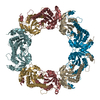
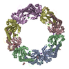
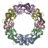
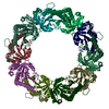
| Title | Circular tandem repeat protein with novel repeat topology and enhanced subunit contact surfaces | ||||||
 Components Components | Circular tendon repeat protein | ||||||
 Keywords Keywords | PEPTIDE BINDING PROTEIN / protein display particles | ||||||
| Biological species | unidentified (others) | ||||||
| Method | ELECTRON MICROSCOPY / single particle reconstruction / cryo EM / Resolution: 6.5 Å | ||||||
 Authors Authors | Shen, B.W. / Stoddard, B.L. | ||||||
| Funding support |  United States, 1items United States, 1items
| ||||||
 Citation Citation | Journal: Commun Biol / Year: 2021 Title: Design of functionalised circular tandem repeat proteins with longer repeat topologies and enhanced subunit contact surfaces. Authors: Jazmine P Hallinan / Lindsey A Doyle / Betty W Shen / Mesfin M Gewe / Brittany Takushi / Madison A Kennedy / Della Friend / James M Roberts / Philip Bradley / Barry L Stoddard / Abstract: Circular tandem repeat proteins ('cTRPs') are de novo designed protein scaffolds (in this and prior studies, based on antiparallel two-helix bundles) that contain repeated protein sequences and ...Circular tandem repeat proteins ('cTRPs') are de novo designed protein scaffolds (in this and prior studies, based on antiparallel two-helix bundles) that contain repeated protein sequences and structural motifs and form closed circular structures. They can display significant stability and solubility, a wide range of sizes, and are useful as protein display particles for biotechnology applications. However, cTRPs also demonstrate inefficient self-assembly from smaller subunits. In this study, we describe a new generation of cTRPs, with longer repeats and increased interaction surfaces, which enhanced the self-assembly of two significantly different sizes of homotrimeric constructs. Finally, we demonstrated functionalization of these constructs with (1) a hexameric array of peptide-binding SH2 domains, and (2) a trimeric array of anti-SARS CoV-2 VHH domains. The latter proved capable of sub-nanomolar binding affinities towards the viral receptor binding domain and potent viral neutralization function. | ||||||
| History |
|
- Structure visualization
| Movie |
 Movie viewer Movie viewer |
|---|---|
| Structure viewer | Molecule: MolmilJmol/JSmol |
- Downloads & links
-Download
| PDBx/mmCIF format | 7rdr.cif.gz | 235.8 KB | Display | PDBx/mmCIF format |
|---|---|---|---|---|
| PDB format | pdb7rdr.ent.gz | 187.6 KB | Display | PDB format |
| PDBx/mmJSON format | 7rdr.json.gz | Tree view | PDBx/mmJSON format | |
| Others |  Other downloads Other downloads |
-Validation report
| Arichive directory | https://data.pdbj.org/pub/pdb/validation_reports/rd/7rdrftp://data.pdbj.org/pub/pdb/validation_reports/rd/7rdr | HTTPS FTP |
|---|
-Related structure data
| Related structure data |  24425MC 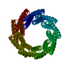 6xr1C 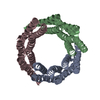 6xr2C M: map data used to model this data C: citing same article ( |
|---|---|
| Similar structure data |


-Links
 PDBj
PDBj
- Assembly
| Deposited unit | 
|
|---|---|
| 1 |
|
-Components
| #1: Protein | Mass: 53141.402 Da / Num. of mol.: 3 Source method: isolated from a genetically manipulated source Source: (gene. exp.) unidentified (others) / Production host:  Has protein modification | Y | |
|---|
-Experimental details
-Experiment
| Experiment | Method: ELECTRON MICROSCOPY |
|---|---|
| EM experiment | Aggregation state: PARTICLE / 3D reconstruction method: single particle reconstruction |
- Sample preparation
| Component | Name: trimer of tendon repeat protein / Type: COMPLEX Details: circular tendon repeats based on de in silico structure design Entity ID: all / Source: RECOMBINANT | |||||||||||||||
|---|---|---|---|---|---|---|---|---|---|---|---|---|---|---|---|---|
| Molecular weight | Value: 0.15 MDa / Experimental value: NO | |||||||||||||||
| Source (natural) | Organism: unidentified (others) | |||||||||||||||
| Source (recombinant) | Organism: | |||||||||||||||
| Buffer solution | pH: 7.5 Details: solution was diluted immediate prior to flash freezing | |||||||||||||||
| Buffer component |
| |||||||||||||||
| Specimen | Conc.: 0.4 mg/ml / Embedding applied: NO / Shadowing applied: NO / Staining applied: NO / Vitrification applied: YES / Details: This sample was more disperse | |||||||||||||||
| Specimen support | Grid material: COPPER / Grid mesh size: 200 divisions/in. / Grid type: Quantifoil R1.2/1.3 | |||||||||||||||
| Vitrification | Instrument: FEI VITROBOT MARK IV / Cryogen name: ETHANE / Humidity: 95 % / Chamber temperature: 298 K / Details: blot for 5 seconds before plunging |
- Electron microscopy imaging
| Microscopy | Model: FEI TECNAI 20 |
|---|---|
| Electron gun | Electron source:  FIELD EMISSION GUN / Accelerating voltage: 200 kV / Illumination mode: SPOT SCAN FIELD EMISSION GUN / Accelerating voltage: 200 kV / Illumination mode: SPOT SCAN |
| Electron lens | Mode: BRIGHT FIELD / Nominal magnification: 38000 X / Nominal defocus max: 2500 nm / Nominal defocus min: 1000 nm / Calibrated defocus min: 1000 nm / Cs: 2.7 mm / C2 aperture diameter: 50 µm / Alignment procedure: COMA FREE |
| Specimen holder | Cryogen: NITROGEN / Specimen holder model: FEI TITAN KRIOS AUTOGRID HOLDER / Temperature (max): 100 K / Temperature (min): 100 K |
| Image recording | Average exposure time: 10 sec. / Electron dose: 40 e/Å2 / Detector mode: COUNTING / Film or detector model: GATAN K2 SUMMIT (4k x 4k) / Num. of grids imaged: 1 / Num. of real images: 420 |
| Image scans | Width: 3710 / Height: 3838 / Used frames/image: 1-50 |
- Processing
| EM software |
| |||||||||||||||||||||||||||||||||||
|---|---|---|---|---|---|---|---|---|---|---|---|---|---|---|---|---|---|---|---|---|---|---|---|---|---|---|---|---|---|---|---|---|---|---|---|---|
| CTF correction | Type: NONE | |||||||||||||||||||||||||||||||||||
| Particle selection | Num. of particles selected: 121850 | |||||||||||||||||||||||||||||||||||
| Symmetry | Point symmetry: C3 (3 fold cyclic) | |||||||||||||||||||||||||||||||||||
| 3D reconstruction | Resolution: 6.5 Å / Resolution method: FSC 0.143 CUT-OFF / Num. of particles: 121400 / Num. of class averages: 4 / Symmetry type: POINT | |||||||||||||||||||||||||||||||||||
| Atomic model building | Protocol: RIGID BODY FIT / Space: REAL |
