[English] 日本語
Yorodumi
- PDB-6xr2: Computationally designed right-handed alpha/alpha homotrimeric to... -

+
Open data


ID or keywords:

Loading...

-
Basic information

Entry
Database: PDB / ID: 6xr2
TitleComputationally designed right-handed alpha/alpha homotrimeric toroid with 3 repeats per subunit
ComponentsdTor_3x57R
KeywordsDE NOVO PROTEIN / Computationally designed / de novo / toroid / helix-turn-helix
Biological speciessynthetic construct (others)
MethodX-RAY DIFFRACTION / MOLECULAR REPLACEMENT / molecular replacement / Resolution: 3.2 Å
AuthorsHallinan, J.P. / Doyle, L. / Bradley, P. / Stoddard, B.L.
Funding support United States, 1items
OrganizationGrant numberCountry
National Institutes of Health/National Institute of General Medical Sciences (NIH/NIGMS)R01GM123378 United States
Citation
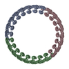
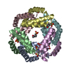
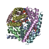
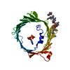
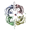
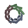
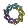
Journal: Commun Biol / Year: 2021
Title: Design of functionalised circular tandem repeat proteins with longer repeat topologies and enhanced subunit contact surfaces.
Authors: Jazmine P Hallinan / Lindsey A Doyle / Betty W Shen / Mesfin M Gewe / Brittany Takushi / Madison A Kennedy / Della Friend / James M Roberts / Philip Bradley / Barry L Stoddard /
Abstract: Circular tandem repeat proteins ('cTRPs') are de novo designed protein scaffolds (in this and prior studies, based on antiparallel two-helix bundles) that contain repeated protein sequences and ...Circular tandem repeat proteins ('cTRPs') are de novo designed protein scaffolds (in this and prior studies, based on antiparallel two-helix bundles) that contain repeated protein sequences and structural motifs and form closed circular structures. They can display significant stability and solubility, a wide range of sizes, and are useful as protein display particles for biotechnology applications. However, cTRPs also demonstrate inefficient self-assembly from smaller subunits. In this study, we describe a new generation of cTRPs, with longer repeats and increased interaction surfaces, which enhanced the self-assembly of two significantly different sizes of homotrimeric constructs. Finally, we demonstrated functionalization of these constructs with (1) a hexameric array of peptide-binding SH2 domains, and (2) a trimeric array of anti-SARS CoV-2 VHH domains. The latter proved capable of sub-nanomolar binding affinities towards the viral receptor binding domain and potent viral neutralization function.
History
DepositionJul 10, 2020Deposition site: RCSB / Processing site: RCSB
Revision 1.0Jul 14, 2021Provider: repository / Type: Initial release
Revision 1.1Nov 10, 2021Group: Database references / Category: citation / citation_author / database_2
Item: _citation.country / _citation.journal_abbrev ..._citation.country / _citation.journal_abbrev / _citation.journal_id_CSD / _citation.journal_id_ISSN / _citation.journal_volume / _citation.page_first / _citation.page_last / _citation.pdbx_database_id_DOI / _citation.pdbx_database_id_PubMed / _citation.title / _citation.year / _database_2.pdbx_DOI / _database_2.pdbx_database_accession
Revision 1.2May 22, 2024Group: Data collection / Category: chem_comp_atom / chem_comp_bond

-
Structure visualization

Structure viewerMolecule:
MolmilJmol/JSmol

Downloads & links

-
Assembly

Deposited unit
A: dTor_3x57R
B: dTor_3x57R
C: dTor_3x57R
D: dTor_3x57R
E: dTor_3x57R
F: dTor_3x57R


Theoretical massNumber of molelcules
Total (without water)113,7096
Polymers113,7096
Non-polymers00
Water23413
1
A: dTor_3x57R
B: dTor_3x57R
C: dTor_3x57R


Theoretical massNumber of molelcules
Total (without water)56,8553
Polymers56,8553
Non-polymers00
Water181
TypeNameSymmetry operationNumber
identity operation1_555x,y,z1
Buried area5720 Å2
ΔGint-48 kcal/mol
Surface area20960 Å2
MethodPISA
2
D: dTor_3x57R
E: dTor_3x57R
F: dTor_3x57R


Theoretical massNumber of molelcules
Total (without water)56,8553
Polymers56,8553
Non-polymers00
Water543
TypeNameSymmetry operationNumber
identity operation1_555x,y,z1
Buried area5440 Å2
ΔGint-48 kcal/mol
Surface area20540 Å2
MethodPISA
Unit cell
Length a, b, c (Å)57.647, 78.930, 112.694
Angle α, β, γ (deg.)90.000, 93.820, 90.000
Int Tables number4
Space group name H-MP1211
Noncrystallographic symmetry (NCS)NCS domain:
IDEns-IDDetails
11(chain A and ((resid 4 and (name N or name...
21(chain B and ((resid 4 and (name N or name...
31(chain C and ((resid 4 and (name N or name...
41(chain D and ((resid 4 and (name O or name...
51(chain E and ((resid 4 and (name O or name...
61(chain F and ((resid 4 and (name O or name...

NCS domain segments:
Dom-IDComponent-IDEns-IDSelection detailsAuth asym-IDAuth seq-ID
111(chain A and ((resid 4 and (name N or name...A0
211(chain B and ((resid 4 and (name N or name...B0
311(chain C and ((resid 4 and (name N or name...C0
411(chain D and ((resid 4 and (name O or name...D0
511(chain E and ((resid 4 and (name O or name...E0
611(chain F and ((resid 4 and (name O or name...F4
621(chain F and ((resid 4 and (name O or name...F2 - 169
631(chain F and ((resid 4 and (name O or name...F2 - 169
641(chain F and ((resid 4 and (name O or name...F2 - 169

-
Components

#1: Protein
dTor_3x57R


Mass: 18951.506 Da / Num. of mol.: 6
Source method: isolated from a genetically manipulated source
Source: (gene. exp.) synthetic construct (others) / Production host: Escherichia coli (E. coli)
#2: Water ChemComp-HOH / water


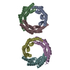
Mass: 18.015 Da / Num. of mol.: 13 / Source method: isolated from a natural source / Formula: H2O

-
Experimental details

-
Experiment

ExperimentMethod: X-RAY DIFFRACTION / Number of used crystals: 1

-
Sample preparation

CrystalDensity Matthews: 2.25 Å3/Da / Density % sol: 45.33 %
Crystal growTemperature: 293 K / Method: vapor diffusion, hanging drop / pH: 4.5
Details: 0.1 M calcium acetate, 0.1 M sodium acetate, pH 4.5, 25% PEG400

-
Data collection

DiffractionMean temperature: 100 K / Serial crystal experiment: N
Diffraction sourceSource: ROTATING ANODE / Type: RIGAKU MICROMAX-007 / Wavelength: 1.54 Å
DetectorType: RIGAKU SATURN 944+ / Detector: CCD / Date: Jan 9, 2017
RadiationProtocol: SINGLE WAVELENGTH / Monochromatic (M) / Laue (L): M / Scattering type: x-ray
Radiation wavelengthWavelength: 1.54 Å / Relative weight: 1
ReflectionResolution: 3.198→50 Å / Num. obs: 14721 / % possible obs: 88 % / Redundancy: 5.5 % / Biso Wilson estimate: 49.76 Å2 / Rmerge(I) obs: 0.221 / Rpim(I) all: 0.097 / Rrim(I) all: 0.243 / Χ2: 1.416 / Net I/σ(I): 5
Reflection shell

Diffraction-ID: 1

Resolution (Å)Redundancy (%)Rmerge(I) obsNum. unique obsCC1/2Rpim(I) allRrim(I) allΧ2% possible all
3.2-3.315.50.69814890.7820.3030.7641.49890
3.31-3.455.50.46414550.8310.2020.5091.47587.9
3.45-3.65.50.4714710.8950.2050.5151.44487.2
3.6-3.795.50.2714660.9280.1180.2961.55589.4
3.79-4.035.30.35814620.9520.1580.3931.55187.7
4.03-4.345.30.25114410.9760.1140.2771.44386.6
4.34-4.785.30.22614240.9780.1010.2491.43385.4
4.78-5.475.30.30614260.9650.1370.3371.4185.5
5.47-6.895.30.20514550.9760.0910.2251.35485.4
6.89-5060.07516320.9960.0320.0821.07394.3

-
Phasing

PhasingMethod: molecular replacement
Phasing MR
Highest resolutionLowest resolution
Rotation7.5 Å30.95 Å
Translation7.5 Å30.95 Å

-
Processing

Software
NameVersionClassification
PHENIX1.1refinement
HKL-2000data scaling
PHASER2.6.0phasing
PDB_EXTRACT3.25data extraction
RefinementMethod to determine structure: MOLECULAR REPLACEMENT / Resolution: 3.2→30.949 Å / SU ML: 0.55 / Cross valid method: THROUGHOUT / σ(F): 1.33 / Phase error: 36.09 / Stereochemistry target values: ML
RfactorNum. reflection% reflection
Rfree0.3292 1472 10.02 %
Rwork0.2426 13218 -
obs0.2514 14690 87.19 %
Solvent computationShrinkage radii: 0.9 Å / VDW probe radii: 1.11 Å / Solvent model: FLAT BULK SOLVENT MODEL
Displacement parametersBiso max: 79.55 Å2 / Biso mean: 30.2152 Å2 / Biso min: 5.46 Å2
Refinement stepCycle: final / Resolution: 3.2→30.949 Å
ProteinNucleic acidLigandSolventTotal
Num. atoms7044 0 0 13 7057
Biso mean---20.2 -
Num. residues----1009
Refine LS restraints
Refine-IDTypeDev idealNumber
X-RAY DIFFRACTIONf_bond_d0.0037060
X-RAY DIFFRACTIONf_angle_d0.7079569
X-RAY DIFFRACTIONf_chiral_restr0.0351193
X-RAY DIFFRACTIONf_plane_restr0.0041291
X-RAY DIFFRACTIONf_dihedral_angle_d18.6534423
Refine LS restraints NCS
Ens-IDDom-IDAuth asym-IDNumberRefine-IDRmsType
11A2839X-RAY DIFFRACTION18.079TORSIONAL
12B2839X-RAY DIFFRACTION18.079TORSIONAL
13C2839X-RAY DIFFRACTION18.079TORSIONAL
14D2839X-RAY DIFFRACTION18.079TORSIONAL
15E2839X-RAY DIFFRACTION18.079TORSIONAL
16F2839X-RAY DIFFRACTION18.079TORSIONAL
LS refinement shell

Refine-ID: X-RAY DIFFRACTION / Rfactor Rfree error: 0

Resolution (Å)Rfactor RfreeNum. reflection RfreeRfactor RworkNum. reflection Rwork% reflection obs (%)
3.2-3.30160.37021160.2811110682
3.3016-3.41950.33811460.2309122188
3.4195-3.55620.37971270.2452120689
3.5562-3.71780.33851380.223121889
3.7178-3.91340.39361330.2508119387
3.9134-4.15810.37991250.2451119186
4.1581-4.47820.30131370.2199118487
4.4782-4.92730.33051340.2265116985
4.9273-5.63650.37231300.2901116584
5.6365-7.08740.29851400.2785119787
7.0874-30.9490.26011460.2261136896

+
About Yorodumi

-
News

-
Feb 9, 2022. New format data for meta-information of EMDB entries

New format data for meta-information of EMDB entries

  • Version 3 of the EMDB header file is now the official format.
  • The previous official version 1.9 will be removed from the archive.

Related info.:EMDB header

External links:wwPDB to switch to version 3 of the EMDB data model

-
Aug 12, 2020. Covid-19 info

Covid-19 info

URL: https://pdbjlvh1.pdbj.org/emnavi/covid19.php

New page: Covid-19 featured information page in EM Navigator.

Related info.:Covid-19 info / Mar 5, 2020. Novel coronavirus structure data

+
Mar 5, 2020. Novel coronavirus structure data

Novel coronavirus structure data

Related info.:Yorodumi Speices / Aug 12, 2020. Covid-19 info

External links:COVID-19 featured content - PDBj / Molecule of the Month (242):Coronavirus Proteases

+
Jan 31, 2019. EMDB accession codes are about to change! (news from PDBe EMDB page)

EMDB accession codes are about to change! (news from PDBe EMDB page)

  • The allocation of 4 digits for EMDB accession codes will soon come to an end. Whilst these codes will remain in use, new EMDB accession codes will include an additional digit and will expand incrementally as the available range of codes is exhausted. The current 4-digit format prefixed with “EMD-” (i.e. EMD-XXXX) will advance to a 5-digit format (i.e. EMD-XXXXX), and so on. It is currently estimated that the 4-digit codes will be depleted around Spring 2019, at which point the 5-digit format will come into force.
  • The EM Navigator/Yorodumi systems omit the EMD- prefix.

Related info.:Q: What is EMD? / ID/Accession-code notation in Yorodumi/EM Navigator

External links:EMDB Accession Codes are Changing Soon! / Contact to PDBj

+
Jul 12, 2017. Major update of PDB

Major update of PDB

  • wwPDB released updated PDB data conforming to the new PDBx/mmCIF dictionary.
  • This is a major update changing the version number from 4 to 5, and with Remediation, in which all the entries are updated.
  • In this update, many items about electron microscopy experimental information are reorganized (e.g. em_software).
  • Now, EM Navigator and Yorodumi are based on the updated data.

External links:wwPDB Remediation / Enriched Model Files Conforming to OneDep Data Standards Now Available in the PDB FTP Archive

-
Yorodumi

Thousand views of thousand structures

  • Yorodumi is a browser for structure data from EMDB, PDB, SASBDB, etc.
  • This page is also the successor to EM Navigator detail page, and also detail information page/front-end page for Omokage search.
  • The word "yorodu" (or yorozu) is an old Japanese word meaning "ten thousand". "mi" (miru) is to see.

Related info.:EMDB / PDB / SASBDB / Comparison of 3 databanks / Yorodumi Search / Aug 31, 2016. New EM Navigator & Yorodumi / Yorodumi Papers / Jmol/JSmol / Function and homology information / Changes in new EM Navigator and Yorodumi

Read more